Human reproductive system

Last updated
Human reproductive system
Anatomy of male and female human genitalia - blanc.png
Internal genitalia of a human female (left) and male (right).
Female and male genitalia.png
External genitalia of an adult female (left) and male (right).
Details
Identifiers
Latin systemata genitalia
TA98 A09.0.00.000
TA2 3467
FMA 7160 75572, 7160
Anatomical terminology

The human reproductive system includes the male reproductive system which functions to produce and deposit sperm; and the female reproductive system which functions to produce egg cells, and to protect and nourish the fetus until birth. Humans have a high level of sexual differentiation. In addition to differences in nearly every reproductive organ, there are numerous differences in typical secondary sex characteristics.

Contents

Human reproduction usually involves internal fertilization by sexual intercourse. In this process, the male inserts his penis into the female's vagina and ejaculates semen, which contains sperm. A small proportion of the sperm pass through the cervix into the uterus, and then into the fallopian tubes for fertilization of the ovum. Only one sperm is required to fertilize the ovum. Upon successful fertilization, the fertilized ovum, or zygote, travels out of the fallopian tube and into the uterus, where it implants in the uterine wall. This marks the beginning of gestation, better known as pregnancy, which continues for around nine months as the fetus develops. When the fetus has developed to a certain point, pregnancy is concluded with childbirth, involving labor. During labor, the muscles of the uterus contract and the cervix dilates over the course of hours, and the baby passes out of the vagina. Human infants are completely dependent on their caregivers, and require high levels of parental care. Infants rely on their caregivers for comfort, cleanliness, and food. Food may be provided by breastfeeding or formula feeding. [1]

Structure

Female

Female reproductive system Female reproductive system lateral 1.png
Female reproductive system

The human female reproductive system is a series of organs primarily located inside the body and around the pelvic region of a female that contribute towards the reproductive process. The human female reproductive system contains three main parts: the vulva, which leads to the vagina, the vaginal opening, to the uterus; the uterus, which holds the developing fetus; and the ovaries, which produce the female's ova. The breasts are involved during the parenting stage of reproduction, but in most classifications they are not considered to be part of the female reproductive system.

The vulva Front and back womans genitalia.jpg
The vulva

The vagina meets the outside at the vulva, which is made up of the labia, clitoris and urethra; during intercourse this area is lubricated by mucus secreted by the Bartholin's glands. The vagina is attached to the uterus through the cervix, while the uterus is attached to the ovaries via the fallopian tubes. Each ovary contains hundreds of egg cells or ova (singular ovum ).

Approximately every 28 days, the pituitary gland releases a hormone that stimulates some of the ova to develop and grow. One ovum is released and it passes through the fallopian tube into the uterus. Hormones produced by the ovaries prepare the uterus to receive the ovum. The lining of the uterus, called the endometrium, and unfertilized ova are shed each cycle through the process of menstruation. If the ovum is fertilized by sperm, it attaches to the endometrium and the fetus develops.

Male

Male reproductive system Male anatomy 1.png
Male reproductive system

The male reproductive system is a series of organs located outside the body and around the pelvis region of a male that contribute towards the reproduction process. The primary direct function of the male reproductive system is to provide the male sperm for fertilization of the ovum.

Penile shrinkage due to low temperatures. The scrotum is in a tense state to regulate testicular temperatures. Flaccid Penis Shrinkage.jpg
Penile shrinkage due to low temperatures. The scrotum is in a tense state to regulate testicular temperatures.

The major reproductive organs of the male can be grouped into three categories. The first category produces and stores sperm (spermatozoa). These are produced in the testicles, which are housed in the temperature-regulating scrotum; immature sperm then travel to the epididymides for development and storage. The second category are the ejaculatory fluid producing glands which include the Cowper's gland (also called bulbourethral gland), seminal vesicles, prostate, and vas deferens. The final category are those used for copulation and deposition of the sperm within the female; these include the penis, urethra, and vas deferens.

Major secondary sexual characteristics include: larger, more muscular stature, deepened voice, facial and body hair, broad shoulders, and development of an Adam's apple. An important sexual hormone of males is androgen, and particularly testosterone.

The testes release a hormone that controls the development of sperm. This hormone is also responsible for the development of physical characteristics in men, such as facial hair and a deep voice.

Development

The development of the reproductive system and the development of the urinary system are closely tied with the development of the human fetus. Despite the differences between them, the adult male and female are determined in early development in the 6th week. The gonads and external genitals are derived from the intermediate mesoderm. [2] The three main fetal precursors of the reproductive organs are the Wolffian duct, the Müllerian ducts, and the gonads. Endocrine hormones are a well-known and critical controlling factor in the normal differentiation of the reproductive system. [3]

The Wolffian duct forms the epididymis, vas deferens, ejaculatory duct, and seminal vesicle in the male reproductive system, but essentially disappears in the female reproductive system. The reverse is true for the Müllerian duct, as it essentially disappears in the male reproductive system and forms the fallopian tubes, uterus, and vagina in the female system. In both sexes, the gonads go on to form the testes and ovaries; because they are derived from the same undeveloped structure, they are considered homologous organs. There are a number of other homologous structures shared between male and female reproductive systems. However, despite the similarity in function of the female fallopian tubes and the male epididymis and vas deferens, they are not homologous but rather analogous structures as they arise from different fetal structures.

Reproduction

Production of gametes

Gametes are produced within the gonads through a process known as gametogenesis. This occurs when certain types of germ cells undergo meiosis to split the normal diploid number of chromosomes (n=46) into haploid cells containing only 23 chromosomes. [4]

Anatomy of the testis Figure 28 01 03.JPG
Anatomy of the testis

In males, this process is known as spermatogenesis, and takes place only after puberty in the seminiferous tubules of the testes. The immature spermatozoa or sperm are then sent to the epididymis, where they gain a tail, enabling motility. Each of the original diploid germ cells or primary spermatocytes forms four functional gametes which is each forever young[ clarification needed ]. The production and survival of sperms require a temperature below the normal core body temperature. Since the scrotum, where the testes is present, is situated outside the body cavity, it provides a temperature about 3 °C below normal body temperature.

In females, gametogenesis is known as oogenesis; this occurs in the ovarian follicles of the ovaries. This process does not produce mature ovum until puberty. In contrast with males, each of the original diploid germ cells or primary oocytes will form only one mature ovum, and three polar bodies which are not capable of fertilization. It has long been understood that in females, unlike males, all the primary oocytes ever found in a female will be created prior to birth, and that the final stages of ova production will then not resume until puberty. [4] However, recent scientific research has challenged that hypothesis. [5] This new research indicates that in at least some species of mammal, oocytes continue to be replenished in females well after birth. [6]

Disease

Like all complex organ systems, the human reproductive system is affected by many diseases. There are four main categories of reproductive diseases in humans. They are:

Rate of sexually transmitted diseases in the U.S Rate of Sexually Transmitted Diseases in the US.svg
Rate of sexually transmitted diseases in the U.S

Specific reproductive diseases are often symptoms of other diseases and disorders, or have multiple, or unknown causes making them difficult to classify. Examples of unclassifiable disorders are Peyronie's disease in males and endometriosis in females. Many congenital conditions cause reproductive abnormalities, but are better known for their other symptoms. These include: Turner syndrome, Klinefelter's syndrome, cystic fibrosis, and Bloom syndrome. [8]

Related Research Articles

<span class="mw-page-title-main">Uterus</span> Female sex organ in mammals

The uterus or womb is the organ in the reproductive system of most female mammals, including humans, that accommodates the embryonic and fetal development of one or more embryos until birth. The uterus is a hormone-responsive sex organ that contains glands in its lining that secrete uterine milk for embryonic nourishment.

<span class="mw-page-title-main">Androgen</span> Any steroid hormone that promotes male characteristics

An androgen is any natural or synthetic steroid hormone that regulates the development and maintenance of male characteristics in vertebrates by binding to androgen receptors. This includes the embryological development of the primary male sex organs, and the development of male secondary sex characteristics at puberty. Androgens are synthesized in the testes, the ovaries, and the adrenal glands.

<span class="mw-page-title-main">Epididymis</span> Tube that connects a testicle to a vas deferens

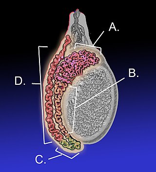
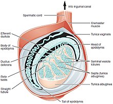
The epididymis is an elongated tubular structure attached to the posterior side of each one of the two male reproductive glands, the testicles. It is a single, narrow, tightly coiled tube in adult humans, 6 to 7 centimetres in length; uncoiled the tube would be approximately 6 m long. It connects the testicle to the vas deferens in the male reproductive system. The epididymis serves as an interconnection between the multiple efferent ducts at the rear of a testicle (proximally), and the vas deferens (distally). Its primary function is the storage, maturation and transport of sperm cells.

<span class="mw-page-title-main">Mesonephric duct</span> Paired organ in mammals

The mesonephric duct, also known as the Wolffian duct, archinephric duct, Leydig's duct or nephric duct, is a paired organ that develops in the early stages of embryonic development in humans and other mammals. It is an important structure that plays a critical role in the formation of male reproductive organs. The duct is named after Caspar Friedrich Wolff, a German physiologist and embryologist who first described it in 1759.

<span class="mw-page-title-main">Female reproductive system</span> Reproductive system of human females

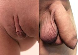
The female reproductive system is made up of the internal and external sex organs that function in the reproduction of new offspring. The human female reproductive system is immature at birth and develops to maturity at puberty to be able to produce gametes, and to carry a fetus to full term. The internal sex organs are the vagina, uterus, fallopian tubes, and ovaries. The female reproductive tract includes the vagina, uterus, and fallopian tubes and is prone to infections. The vagina allows for sexual intercourse and childbirth, and is connected to the uterus at the cervix. The uterus or womb accommodates the embryo, which develops into the fetus. The uterus also produces secretions, which help the transit of sperm to the fallopian tubes, where sperm fertilize ova produced by the ovaries. The external sex organs are also known as the genitals and these are the organs of the vulva including the labia, clitoris, and vaginal opening.

<span class="mw-page-title-main">Paramesonephric duct</span> Paired ducts in the embryo in the primitive urogenital structures

The paramesonephric ducts are paired ducts of the embryo in the female reproductive system that run down the lateral sides of the genital ridge and terminate at the sinus tubercle in the primitive urogenital sinus. In the female, they will develop to form the fallopian tubes, uterus, cervix, and the upper one-third of the vagina.

<span class="mw-page-title-main">Persistent Müllerian duct syndrome</span> Medical condition

Persistent Müllerian duct syndrome (PMDS) is the presence of Müllerian duct derivatives in what would be considered a genetically and otherwise physically normal male animal by typical human based standards. In humans, PMDS typically is due to an autosomal recessive congenital disorder and is considered by some to be a form of pseudohermaphroditism due to the presence of Müllerian derivatives. PMDS can also present in non-human animals.

The development of the urinary system begins during prenatal development, and relates to the development of the urogenital system – both the organs of the urinary system and the sex organs of the reproductive system. The development continues as a part of sexual differentiation.

Reproductive biology includes both sexual and asexual reproduction.

<span class="mw-page-title-main">Male reproductive system</span> Reproductive system of the human male

The male reproductive system consists of a number of sex organs that play a role in the process of human reproduction. These organs are located on the outside of the body, and within the pelvis.

<span class="mw-page-title-main">Human reproduction</span> Procreative biological processes of humanity

Human reproduction is sexual reproduction that results in human fertilization to produce a human offspring. It typically involves sexual intercourse between a sexually mature human male and female. During sexual intercourse, the interaction between the male and female reproductive systems results in fertilization of the ovum by the sperm to form a zygote. While normal cells contain 46 chromosomes, gamete cells only contain 23 single chromosomes, and it is when these two cells merge into one zygote cell that genetic recombination occurs and the new zygote contains 23 chromosomes from each parent, giving it 46 chromosomes. The zygote then undergoes a defined development process that is known as human embryogenesis, and this starts the typical 9-month gestation period that is followed by childbirth. The fertilization of the ovum may be achieved by artificial insemination methods, which do not involve sexual intercourse. Assisted reproductive technology also exists.

A reproductive system disease is any disease of the human reproductive system.

<span class="mw-page-title-main">Sex cords</span> Structures that develop from the genital ridges that further differentiate based on an embryos sex

Sex cords are embryonic structures which eventually will give rise (differentiate) to the adult gonads. They are formed from the genital ridges - which will develop into the gonads - in the first 2 months of gestation which depending on the sex of the embryo will give rise to male or female sex cords. These epithelial cells penetrate and invade the underlying mesenchyme to form the primitive sex cords. This occurs shortly before and during the arrival of the primordial germ cells (PGCs) to the paired genital ridges. If there is a Y chromosome present, testicular cords will develop via the Sry gene : repressing the female sex cord genes and activating the male. If there is no Y chromosome present the opposite will occur, developing ovarian cords. Prior to giving rise to sex cords, both XX and XY embryos have Müllerian ducts and Wolffian ducts. One of these structures will be repressed to induce the other to further differentiate into the external genitalia.

<span class="mw-page-title-main">Sexual differentiation in humans</span> Process of development of sex differences in humans

Sexual differentiation in humans is the process of development of sex differences in humans. It is defined as the development of phenotypic structures consequent to the action of hormones produced following gonadal determination. Sexual differentiation includes development of different genitalia and the internal genital tracts and body hair plays a role in sex identification.

The development of the reproductive system is the part of embryonic growth that results in the sex organs and contributes to sexual differentiation. Due to its large overlap with development of the urinary system, the two systems are typically described together as the genitourinary system.

The reproductive system of an organism, also known as the genital system, is the biological system made up of all the anatomical organs involved in sexual reproduction. Many non-living substances such as fluids, hormones, and pheromones are also important accessories to the reproductive system. Unlike most organ systems, the sexes of differentiated species often have significant differences. These differences allow for a combination of genetic material between two individuals, which allows for the possibility of greater genetic fitness of the offspring.

<span class="mw-page-title-main">Reproductive system of gastropods</span>

The reproductive system of gastropods varies greatly from one group to another within this very large and diverse taxonomic class of animals. Their reproductive strategies also vary greatly.

<span class="mw-page-title-main">Fallopian tube</span> Tubes in the human female reproductive system

The fallopian tubes, also known as uterine tubes, oviducts or salpinges, are paired tubes in the human female body that stretch from the ovaries to the uterus. The fallopian tubes are part of the female reproductive system. In other vertebrates, they are only called oviducts.

<span class="mw-page-title-main">Mammalian reproduction</span> Most mammals are viviparous, giving birth to live young

Most mammals are viviparous, giving birth to live young. However, the five species of monotreme, the platypuses and the echidnas, lay eggs. The monotremes have a sex determination system different from that of most other mammals. In particular, the sex chromosomes of a platypus are more like those of a chicken than those of a therian mammal.

Heterocrine glands are the glands which function as both exocrine gland and endocrine gland. These glands exhibit a unique and diverse secretory function encompassing the release of proteins and non-proteinaceous compounds, endocrine and exocrine secretions into both the bloodstream and ducts respectively, thereby bridging the realms of internal and external communication within the body. This duality allows them to serve crucial roles in regulating various physiological processes and maintaining homeostasis. These include the gonads, pancreas and salivary glands.

References

  1. Sexual Reproduction in Humans. Archived 2018-02-17 at the Wayback Machine 2006. John W. Kimball. Kimball's Biology Pages, and online textbook.
  2. Rey, Rodolfo; Josso, Nathalie; Racine, Chrystèle (2000), Feingold, Kenneth R.; Anawalt, Bradley; Blackman, Marc R.; Boyce, Alison (eds.), "Sexual Differentiation", Endotext, South Dartmouth (MA): MDText.com, Inc., PMID   25905232 , retrieved 2023-12-19
  3. EDRI Federal Project Inventory: Cellular and Molecular Mechanisms of Abnormal Reproductive Development Archived 2008-12-06 at the Wayback Machine US EPA. Dr. William R. Kelce. 2006.
  4. 1 2 Development of sex cells Archived 2008-07-05 at the Wayback Machine in Reproductive system, Body Guide. Adam.
  5. Tilly JL, Niikura Y, Rueda BR (August 2008). "The Current Status of Evidence for and Against Postnatal Oogenesis in Mammals: A Case of Ovarian Optimism Versus Pessimism?". Biol. Reprod. 80 (1): 2–12. doi:10.1095/biolreprod.108.069088. PMC   2804806 . PMID   18753611.
  6. Johnson J, Canning J, Kaneko T, Pru JK, Tilly JL (March 2004). "Germline stem cells and follicular renewal in the postnatal mammalian ovary". Nature. 428 (6979): 145–50. Bibcode:2004Natur.428..145J. doi:10.1038/nature02316. PMID   15014492. S2CID   1124530.
  7. Jensen, Christian Fuglesang S.; Østergren, Peter; Dupree, James M.; Ohl, Dana A.; Sønksen, Jens; Fode, Mikkel (September 2017). "Varicocele and male infertility". Nature Reviews Urology. 14 (9): 523–533. doi:10.1038/nrurol.2017.98. ISSN   1759-4820. PMID   28675168. S2CID   19357838. Archived from the original on 2022-08-27. Retrieved 2022-08-27.
  8. Genetic Conditions > Reproductive system. Archived 2008-12-04 at the Wayback Machine 2007. Genetics Home Reference. U.S. National Library of Medicine.