Pantothenate kinase

Last updated
Pantothenate kinase
Identifiers
EC no. 2.7.1.33
CAS no. 9026-48-6
Databases
IntEnz IntEnz view
BRENDA BRENDA entry
ExPASy NiceZyme view
KEGG KEGG entry
MetaCyc metabolic pathway
PRIAM profile
PDB structures RCSB PDB PDBe PDBsum
Search
PMC articles
PubMed articles
NCBI proteins

Pantothenate kinase (EC 2.7.1.33, PanK; CoaA) is the first enzyme in the Coenzyme A (CoA) biosynthetic pathway. It phosphorylates pantothenate (vitamin B5) to form 4'-phosphopantothenate at the expense of a molecule of adenosine triphosphate (ATP). It is the rate-limiting step in the biosynthesis of CoA. [1] [2]

Contents

Mechanism os pantothenate kinase.png

CoA is a necessary cofactor in all living organisms. It acts as the major acyl group carrier in many important cellular processes, such as the citric acid cycle (tricarboxylic acid cycle) and fatty acid metabolism. Consequently, pantothenate kinase is a key regulatory enzyme in the CoA biosynthetic pathway. [3]

Types

Three distinct types of PanK has been identified - PanK-I (found in bacteria), PanK-II (mainly found in eukaryotes, but also in the Staphylococci ) and PanK-III, also known as CoaX (found in bacteria). Eukaryotic PanK-II enzymes often occur as different isoforms, such as PanK1, PanK2, PanK3 and PanK4. In humans, multiple PanK isoforms are expressed by four genes. PANK1 gene encodes the PanK1α and PanK1β forms, and PANK2 and PANK3 encode PanK2 and PanK3, respectively. [4] The four major isoforms found in mammals have different subcellular localizations. PanK1α is nuclear, while PanK1β and PanK3 are cytosolic. In mice, PanK2 is also cytosolic, while in humans, this enzyme is mitochondrial and nuclear. [5] The tissue distribution of these isoforms also varies. In mouse models, PanK1 is the predominant species in the heart, liver and brown adipose tissue, along with the kidneys. PanK2 and PanK3 are more prominent in the brain and skeletal muscle, and PanK3 is particularly high in the intestines and white adipose tissue. [6]

Structure

PanK-II

Fig. 1 Dimer structure of PanK-II Human PanK-II structure.png
Fig. 1 Dimer structure of PanK-II

PanK-II contains two protein domains, as illustrated in Figure 1. The A domain and A' domain each has a glycine-rich loop (sequence GXXXXGKS; P loop) that is characteristic of nucleotide-binding sites; this is where ATP is assumed to bind. [7] located between residues 95 and 102 on the A domain

The two ATP binding sites display cooperative behavior. The dimerization interface consists of two long helices, one from each monomer, that interact with each other. The C-terminal ends of the helices are held together by van der Waals interactions between valine and methionine residues of each monomer. The middle of the helices is attached by hydrogen bonds between asparagine residues. At the N-terminal end, each helix widens and forms a four-helix bundle with two shorter helices. This bundle consists of a hydrophobic core formed by non-polar residues that utilize van der Waals forces to further stabilize the dimer. [4]

In the active site, pantothenate is oriented by hydrogen bonds between pantothenate and the side chains of aspartate, tyrosine, histidine, tyrosine, and asparagine residues. [8] Asparagine, histidine, and arginine residues are involved in catalysis.

Human PanK-II isoforms PanK1α, PanK1β, PanK2, and PanK3 have a common, highly homologous catalytic core of approximately 355 residues. [4] PanK1α and PanK1β are both encoded by the PANK1 gene and have the same catalytic domain of 363 amino acids, encoded by exons 2 through 7. The PanK1α transcript starts with exon 1α that encodes a 184-residue regulatory domain at the N-terminus. This region allows for feedback inhibition by free CoA and acyl-CoA and regulation by acetyl-CoA and malonyl-CoA. On the other hand, the PanK1β transcript starts with exon 1β, which produces a 10-residue N-terminus that does not include a feedback regulatory domain. [9]

PanK-III

Fig. 2 Dimer structure of PanK-III PanK-III.png
Fig. 2 Dimer structure of PanK-III

PanK-III also contains two protein domains, and the key catalytic residues of PanK-II are conserved. The monomer units of PanK-II and PanK-III are virtually identical, but they have distinctly different dimer assemblies. A study between the structures of Staphylococcus aureus type II and the Pseudomonas aeruginosa type III demonstrate that the PanK-II monomer has a loop region that is absent from the PanK-III monomer, and the PanK-III monomer has a loop region that is absent from the PanK-II monomer. [10] This minor variation has a crucial difference on the dimerization interface in which the helices of the PanK-II dimer coil around one another and the helices of the PanK-III dimer interact at a 70° angle (Figure 2). [11]

As a result of this difference in dimerization interface between PanK-II and PanK-III, the conformations of the substrate binding sites for ATP and pantothenate are also distinct. [12] [13]

Catalytic Mechanism

Fig. 3 Proposed catalytic mechanism for PanK-II Pantothenate Kinase mechanism.png
Fig. 3 Proposed catalytic mechanism for PanK-II

PanK-II

A proposed mechanism of the phosphoryl transfer reaction of PanK-II is a concerted mechanism with a dissociative transition state.

First, the ATP binds at the binding groove created by residues of the P loop and nearby residues. Here, the conserved lysine (Lys-101) is the key residue required for ATP binding. [14] [15] Additionally, the side chains of residues Lys-101, Ser-102, Glu-199, and Arg-243 orient the nucleotide in the binding groove. The pantothenate is bound and oriented by forming hydrogen bond interactions with residues Asp-127, Tyr-240, Asn-282, Tyr-175, and His-177. [8] When both ATP and pantothenate are bound, Asp-127 deprotonates the C1 hydroxyl group of pantothenate. The oxygen from the pantothenate then attacks the γ-phosphate of the bound ATP. Here, charge stabilization of β- and γ-phosphate groups is achieved by Arg-243, Lys-101, and a coordinated Mg2+ ion. [16] In this concerted mechanism, the planar phosphorane of the γ-phosphate is transferred in-line to the attacking oxygen of pantothenate. [8] Finally, 4'-phosphopantothenate dissociates from PanK, followed by ADP.

Regulation of pantothenate kinase

PanK-II

The regulation of pantothenate kinase is essential to controlling the intracellular CoA concentration. [17] Pantothenate kinase is regulated via feedback inhibition by CoA and its thioesters (i.e., acetyl-CoA, malonyl-CoA). [18] Inhibition of the human isoforms of PanK by acetyl-CoA varies dramatically. PanK1β is inhibited the least strongly, with an IC50 value of around 5 μM, while PanK2 is the most strongly inhibited, with an IC50 of around 0.1 μM. [6]

CoA inhibits PanK activity by competitively binding to the ATP binding site and preventing ATP binding to Lys-101. [14] [15] Although CoA binds at the same site as ATP, they bind in distinct orientations, and their adenine moieties interact with the enzyme with nonoverlapping sets of residues. His-177, Phe-247, and Arg-106 are necessary for CoA recognition but not for ATP, and while Asn-43 and His-307 interact with the adenine base of ATP, His-177 and Phe-247 interact with the adenine base of CoA. [16] Both molecules use Lys-101 to neutralize the charge on their respective phosphodiesters.

Nonesterified CoA has more potent inhibition than its thioesters. This phenomenon is best explained by the tight fit of the thiol group with the surrounding aromatic residues, Phe-244, Phe-259, Tyr-262, and Phe-252. Free CoA has an optimal fit, but when an acyl group is attached to CoA, the steric hindrance makes it difficult for the thioester to interact with Phe-252. Thus, the inhibition by thioesters is less effective than that by nonesterified CoA. [16]

Deletion of PanK1 disrupts metabolic pathways, including fatty acid oxidation and gluconeogenesis. PanK1-/- mouse models in a fasted state show impaired gluconeogenesis, indicating that this pathway is disrupted. In addition, CoA levels decrease significantly between PanK1-/- and wild-type mice. This reduction in CoA also appears to correlate with a disruption in fatty acid oxidation. Higher levels of long-chain acyl-carnitines were observed in PanK1-/- mice, indicating a lower capability for fatty acid oxidation in these mice. [19]

PanK2 Regulation PanK Figure.png
PanK2 Regulation

Because PanK2 is so strongly inhibited by acetyl-CoA, an abundant metabolite in the mitochondria, this enzyme likely would not be active under physiological conditions without activators. [6] Palmitoyl-carnitine and other long-chain acyl-carnitines can reverse acetyl-CoA inhibition and can activate PanK2 without acetyl-CoA present. Palmitoyl-carnitine is competitive with acetyl-CoA. [20] The activation of PanK2 by palmitoyl-carnitine and other long-chain acyl-carnitines sheds light on the regulatory pathways of this enzyme: Under normal conditions, PanK2 is likely inhibited by high levels of acetyl-CoA. Without CoA production, fatty acid oxidation decreases, leading to an increase in long-chain acyl-carnitines. [19] These acyl-carnitines can then reduce inhibition by acetyl-CoA, activating PanK2 and increasing CoA biosynthesis. PanK3 is also activated by palmitoyl-carnitine and other long-chain acyl-carnitines, including oleoyl-carnitine. [21]

PanK-III

The regulation outlined above corresponds to PanK-II. PanK-III is resistant to feedback inhibition. [10] [12] [13]

Genes

In humans:

The PANK2 gene encodes for PanK2, which regulates the formation of CoA in mitochondria, the cell’s energy-producing centers. [22] PANK2 mutation is the cause of Pantothenate kinase-associated neurodegeneration (PKAN), formerly called Hallervorden-Spatz syndrome. This rare disease presents with profound dystonia, spasticity and is often fatal.

There are many mutations in PanK2 that lead to PKAN. In a survey of several common mutations, it was found that several of these mutations did not cause a major loss in the catalytic activity of PanK2, indicating that loss of catalytic function of this enzyme is not fully responsible for this disease. [23]

Related Research Articles

<span class="mw-page-title-main">Pantothenic acid</span> Chemical compound

Pantothenic acid (vitamin B5) is a B vitamin and an essential nutrient. All animals need pantothenic acid in order to synthesize coenzyme A (CoA)—essential for metabolizing fatty acid—and to synthesize and metabolize proteins, carbohydrates, and fats.

<span class="mw-page-title-main">Coenzyme A</span> Coenzyme, notable for its synthesis and oxidation role

Coenzyme A (CoA, SHCoA, CoASH) is a coenzyme, notable for its role in the synthesis and oxidation of fatty acids, and the oxidation of pyruvate in the citric acid cycle. All genomes sequenced to date encode enzymes that use coenzyme A as a substrate, and around 4% of cellular enzymes use it (or a thioester) as a substrate. In humans, CoA biosynthesis requires cysteine, pantothenate (vitamin B5), and adenosine triphosphate (ATP).

<span class="mw-page-title-main">Carnitine</span> Amino acid active in mitochondria

Carnitine is a quaternary ammonium compound involved in metabolism in most mammals, plants, and some bacteria. In support of energy metabolism, carnitine transports long-chain fatty acids from the cytosol into mitochondria to be oxidized for free energy production, and also participates in removing products of metabolism from cells. Given its key metabolic roles, carnitine is concentrated in tissues like skeletal and cardiac muscle that metabolize fatty acids as an energy source. Generally individuals, including strict vegetarians, synthesize enough L-carnitine in vivo.

<span class="mw-page-title-main">Acetyl-CoA carboxylase</span> Enzyme that regulates the metabolism of fatty acids

Acetyl-CoA carboxylase (ACC) is a biotin-dependent enzyme that catalyzes the irreversible carboxylation of acetyl-CoA to produce malonyl-CoA through its two catalytic activities, biotin carboxylase (BC) and carboxyltransferase (CT). ACC is a multi-subunit enzyme in most prokaryotes and in the chloroplasts of most plants and algae, whereas it is a large, multi-domain enzyme in the cytoplasm of most eukaryotes. The most important function of ACC is to provide the malonyl-CoA substrate for the biosynthesis of fatty acids. The activity of ACC can be controlled at the transcriptional level as well as by small molecule modulators and covalent modification. The human genome contains the genes for two different ACCs—ACACA and ACACB.

<span class="mw-page-title-main">Glucose-6-phosphate dehydrogenase</span> Enzyme involved in the production of energy by cells

Glucose-6-phosphate dehydrogenase (G6PD or G6PDH) (EC 1.1.1.49) is a cytosolic enzyme that catalyzes the chemical reaction

Pantothenate kinase-associated neurodegeneration (PKAN), formerly called Hallervorden–Spatz syndrome, is a genetic degenerative disease of the brain that can lead to parkinsonism, dystonia, dementia, and ultimately death. Neurodegeneration in PKAN is accompanied by an excess of iron that progressively builds up in the brain.

Fatty acid degradation is the process in which fatty acids are broken down into their metabolites, in the end generating acetyl-CoA, the entry molecule for the citric acid cycle, the main energy supply of living organisms, including bacteria and animals. It includes three major steps:

<span class="mw-page-title-main">Long-chain-fatty-acid—CoA ligase</span> Class of enzymes

The long chain fatty acyl-CoA ligase is an enzyme of the ligase family that activates the oxidation of complex fatty acids. Long chain fatty acyl-CoA synthetase catalyzes the formation of fatty acyl-CoA by a two-step process proceeding through an adenylated intermediate. The enzyme catalyzes the following reaction,

<span class="mw-page-title-main">Carnitine palmitoyltransferase I</span> Protein-coding gene in the species Homo sapiens

Carnitine palmitoyltransferase I (CPT1) also known as carnitine acyltransferase I, CPTI, CAT1, CoA:carnitine acyl transferase (CCAT), or palmitoylCoA transferase I, is a mitochondrial enzyme responsible for the formation of acyl carnitines by catalyzing the transfer of the acyl group of a long-chain fatty acyl-CoA from coenzyme A to l-carnitine. The product is often Palmitoylcarnitine, but other fatty acids may also be substrates. It is part of a family of enzymes called carnitine acyltransferases. This "preparation" allows for subsequent movement of the acyl carnitine from the cytosol into the intermembrane space of mitochondria.

<span class="mw-page-title-main">ACAT1</span> Protein-coding gene in the species Homo sapiens

Acetyl-CoA acetyltransferase, mitochondrial, also known as acetoacetyl-CoA thiolase, is an enzyme that in humans is encoded by the ACAT1 gene.

<span class="mw-page-title-main">ATP citrate synthase</span> Class of enzymes

ATP citrate synthase (also ATP citrate lyase (ACLY)) is an enzyme that in animals represents an important step in fatty acid biosynthesis. By converting citrate to acetyl-CoA, the enzyme links carbohydrate metabolism, which yields citrate as an intermediate, with fatty acid biosynthesis, which consumes acetyl-CoA. In plants, ATP citrate lyase generates cytosolic acetyl-CoA precursors of thousands of specialized metabolites, including waxes, sterols, and polyketides.

<span class="mw-page-title-main">Carnitine O-acetyltransferase</span> Enzyme

Carnitine O-acetyltransferase also called carnitine acetyltransferase is an enzyme that encoded by the CRAT gene that catalyzes the chemical reaction

<span class="mw-page-title-main">Carnitine O-octanoyltransferase</span>

Carnitine O-octanoyltransferase is a member of the transferase family, more specifically a carnitine acyltransferase, a type of enzyme which catalyzes the transfer of acyl groups from acyl-CoAs to carnitine, generating CoA and an acyl-carnitine. The systematic name of this enzyme is octanoyl-CoA:L-carnitine O-octanoyltransferase. Other names in common use include medium-chain/long-chain carnitine acyltransferase, carnitine medium-chain acyltransferase, easily solubilized mitochondrial carnitine palmitoyltransferase, and overt mitochondrial carnitine palmitoyltransferase. Specifically, CROT catalyzes the chemical reaction:

<span class="mw-page-title-main">PANK2 (gene)</span> Protein-coding gene in the species Homo sapiens

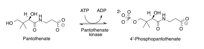
Pantothenate kinase 2, mitochondrial is an enzyme that in humans is encoded by the PANK2 gene.

<span class="mw-page-title-main">ACOT11</span> Protein-coding gene in the species Homo sapiens

Acyl-coenzyme A thioesterase 11 also known as StAR-related lipid transfer protein 14 (STARD14) is an enzyme that in humans is encoded by the ACOT11 gene. This gene encodes a protein with acyl-CoA thioesterase activity towards medium (C12) and long-chain (C18) fatty acyl-CoA substrates which relies on its StAR-related lipid transfer domain. Expression of a similar murine protein in brown adipose tissue is induced by cold exposure and repressed by warmth. Expression of the mouse protein has been associated with obesity, with higher expression found in obesity-resistant mice compared with obesity-prone mice. Alternative splicing results in two transcript variants encoding different isoforms.

<span class="mw-page-title-main">PANK4</span> Protein-coding gene in the species Homo sapiens

Pantothenate kinase 4 is an enzyme that in humans is encoded by the PANK4 gene.

<span class="mw-page-title-main">PANK1</span> Protein-coding gene in the species Homo sapiens

Pantothenate kinase 1 is an enzyme that in humans is encoded by the PANK1 gene.

<span class="mw-page-title-main">ACOT1</span> Protein-coding gene in the species Homo sapiens

Acyl-CoA thioesterase 1 is a protein that in humans is encoded by the ACOT1 gene.

<span class="mw-page-title-main">Bump and hole</span>

The bump-and-hole method is a tool in chemical genetics for studying a specific isoform in a protein family without perturbing the other members of the family. The unattainability of isoform-selective inhibition due to structural homology in protein families is a major challenge of chemical genetics. With the bump-and-hole approach, a protein–ligand interface is engineered to achieve selectivity through steric complementarity while maintaining biochemical competence and orthogonality to the wild type pair. Typically, a "bumped" ligand/inhibitor analog is designed to bind a corresponding "hole-modified" protein. Bumped ligands are commonly bulkier derivatives of a cofactor of the target protein. Hole-modified proteins are recombinantly expressed with an amino acid substitution from a larger to smaller residue, e.g. glycine or alanine, at the cofactor binding site. The designed ligand/inhibitor has specificity for the engineered protein due to steric complementarity, but not the native counterpart due to steric interference.

Andrimid is an antibiotic natural product that is produced by the marine bacterium Vibrio coralliilyticus. Andrimid is an inhibitor of fatty acid biosynthesis by blocking the carboxyl transfer reaction of acetyl-CoA carboxylase (ACC).

References

  1. Robishaw JD, Berkich D, Neely JR (September 1982). "Rate-limiting step and control of coenzyme A synthesis in cardiac muscle". The Journal of Biological Chemistry. 257 (18): 10967–72. doi: 10.1016/S0021-9258(18)33918-8 . PMID   7107640.
  2. 1 2 Yang K, Eyobo Y, Brand LA, Martynowski D, Tomchick D, Strauss E, Zhang H (August 2006). "Crystal structure of a type III pantothenate kinase: insight into the mechanism of an essential coenzyme A biosynthetic enzyme universally distributed in bacteria". Journal of Bacteriology. 188 (15): 5532–40. doi:10.1128/JB.00469-06. PMC   1540032 . PMID   16855243.
  3. Leonardi R, Zhang YM, Rock CO, Jackowski S (2005-03-01). "Coenzyme A: back in action". Progress in Lipid Research. 44 (2–3): 125–53. doi:10.1016/j.plipres.2005.04.001. PMID   15893380.
  4. 1 2 3 Hong BS, Senisterra G, Rabeh WM, Vedadi M, Leonardi R, Zhang YM, Rock CO, Jackowski S, Park HW (September 2007). "Crystal structures of human pantothenate kinases. Insights into allosteric regulation and mutations linked to a neurodegeneration disorder". The Journal of Biological Chemistry. 282 (38): 27984–93. doi: 10.1074/jbc.M701915200 . PMID   17631502.
  5. Alfonso-Pecchio, Adolfo; Garcia, Matthew; Leonardi, Roberta; Jackowski, Suzanne (2012). "Compartmentalization of mammalian pantothenate kinases". PLOS ONE. 7 (11): e49509. doi: 10.1371/journal.pone.0049509 . ISSN   1932-6203. PMC   3496714 . PMID   23152917.
  6. 1 2 3 Dansie, Lorraine E.; Reeves, Stacy; Miller, Karen; Zano, Stephen P.; Frank, Matthew; Pate, Caroline; Wang, Jina; Jackowski, Suzanne (August 2014). "Physiological roles of the pantothenate kinases". Biochemical Society Transactions. 42 (4): 1033–1036. doi:10.1042/BST20140096. ISSN   1470-8752. PMC   4948118 . PMID   25109998.
  7. Saraste M, Sibbald PR, Wittinghofer A (November 1990). "The P-loop--a common motif in ATP- and GTP-binding proteins". Trends in Biochemical Sciences. 15 (11): 430–4. doi:10.1016/0968-0004(90)90281-F. PMID   2126155.
  8. 1 2 3 Ivey RA, Zhang YM, Virga KG, Hevener K, Lee RE, Rock CO, Jackowski S, Park HW (August 2004). "The structure of the pantothenate kinase.ADP.pantothenate ternary complex reveals the relationship between the binding sites for substrate, allosteric regulator, and antimetabolites". The Journal of Biological Chemistry. 279 (34): 35622–9. doi: 10.1074/jbc.M403152200 . PMID   15136582.
  9. Rock CO, Karim MA, Zhang YM, Jackowski S (2002). "The murine pantothenate kinase (Pank1) gene encodes two differentially regulated pantothenate kinase isozymes". Gene. 291 (1–2): 35–43. doi:10.1016/S0378-1119(02)00564-4. PMID   12095677.
  10. 1 2 Yang K, Strauss E, Huerta C, Zhang H (February 2008). "Structural basis for substrate binding and the catalytic mechanism of type III pantothenate kinase". Biochemistry. 47 (5): 1369–80. doi:10.1021/bi7018578. PMID   18186650.
  11. Hong BS, Yun MK, Zhang YM, Chohnan S, Rock CO, White SW, Jackowski S, Park HW, Leonardi R (August 2006). "Prokaryotic type II and type III pantothenate kinases: The same monomer fold creates dimers with distinct catalytic properties". Structure. 14 (8): 1251–61. doi: 10.1016/j.str.2006.06.008 . PMID   16905099.
  12. 1 2 Brand LA, Strauss E (May 2005). "Characterization of a new pantothenate kinase isoform from Helicobacter pylori". The Journal of Biological Chemistry. 280 (21): 20185–8. doi: 10.1074/jbc.C500044200 . PMID   15795230.
  13. 1 2 Choudhry AE, Mandichak TL, Broskey JP, Egolf RW, Kinsland C, Begley TP, Seefeld MA, Ku TW, Brown JR, Zalacain M, Ratnam K (June 2003). "Inhibitors of pantothenate kinase: novel antibiotics for staphylococcal infections". Antimicrobial Agents and Chemotherapy. 47 (6): 2051–5. doi:10.1128/AAC.47.6.2051-2055.2003. PMC   155856 . PMID   12760898.
  14. 1 2 Song WJ, Jackowski S (October 1994). "Kinetics and regulation of pantothenate kinase from Escherichia coli". The Journal of Biological Chemistry. 269 (43): 27051–8. doi: 10.1016/S0021-9258(18)47124-4 . PMID   7929447.
  15. 1 2 Song WJ, Jackowski S (October 1992). "Cloning, sequencing, and expression of the pantothenate kinase (coaA) gene of Escherichia coli". Journal of Bacteriology. 174 (20): 6411–7. doi:10.1128/jb.174.20.6411-6417.1992. PMC   207592 . PMID   1328157.
  16. 1 2 3 Yun M, Park CG, Kim JY, Rock CO, Jackowski S, Park HW (September 2000). "Structural basis for the feedback regulation of Escherichia coli pantothenate kinase by coenzyme A". The Journal of Biological Chemistry. 275 (36): 28093–9. doi: 10.1074/jbc.M003190200 . PMID   10862768.
  17. Jackowski S, Rock CO (December 1981). "Regulation of coenzyme A biosynthesis". Journal of Bacteriology. 148 (3): 926–32. doi:10.1128/jb.148.3.926-932.1981. PMC   216294 . PMID   6796563.
  18. Rock CO, Park HW, Jackowski S (June 2003). "Role of feedback regulation of pantothenate kinase (CoaA) in control of coenzyme A levels in Escherichia coli". Journal of Bacteriology. 185 (11): 3410–5. doi:10.1128/JB.185.11.3410-3415.2003. PMC   155388 . PMID   12754240.
  19. 1 2 Leonardi, Roberta; Rehg, Jerold E.; Rock, Charles O.; Jackowski, Suzanne (2010-06-14). "Pantothenate kinase 1 is required to support the metabolic transition from the fed to the fasted state". PLOS ONE. 5 (6): e11107. Bibcode:2010PLoSO...511107L. doi: 10.1371/journal.pone.0011107 . ISSN   1932-6203. PMC   2885419 . PMID   20559429.
  20. Leonardi, Roberta; Rock, Charles O.; Jackowski, Suzanne; Zhang, Yong-Mei (2007-01-30). "Activation of human mitochondrial pantothenate kinase 2 by palmitoylcarnitine". Proceedings of the National Academy of Sciences. 104 (5): 1494–1499. doi: 10.1073/pnas.0607621104 . ISSN   0027-8424. PMC   1785270 . PMID   17242360.
  21. Leonardi, Roberta; Zhang, Yong-Mei; Yun, Mi-Kyung; Zhou, Ruobing; Zeng, Fu-Yue; Lin, Wenwei; Cui, Jimmy; Chen, Taosheng; Rock, Charles O.; White, Stephen W.; Jackowski, Suzanne (2010-08-27). "Modulation of Pantothenate Kinase 3 Activity by Small Molecules that Interact with the Substrate/Allosteric Regulatory Domain". Chemistry & Biology. 17 (8): 892–902. doi:10.1016/j.chembiol.2010.06.006. ISSN   1074-5521. PMC   2929395 . PMID   20797618.
  22. "PANK2 gene". Genetics Home Reference. 2016-02-22. Retrieved 2016-02-29.
  23. Zhang, Yong-Mei; Rock, Charles O.; Jackowski, Suzanne (2006-01-06). "Biochemical properties of human pantothenate kinase 2 isoforms and mutations linked to pantothenate kinase-associated neurodegeneration". The Journal of Biological Chemistry. 281 (1): 107–114. doi: 10.1074/jbc.M508825200 . ISSN   0021-9258. PMID   16272150.