Complement factor I

Last updated
CFI
Available structures
PDB Ortholog search: PDBe RCSB
Identifiers
Aliases CFI , AHUS3, ARMD13, C3BINA, C3b-INA, FI, IF, KAF, complement factor I
External IDs OMIM: 217030 MGI: 105937 HomoloGene: 171 GeneCards: CFI
Orthologs
SpeciesHumanMouse
Entrez
Ensembl
UniProt
RefSeq (mRNA)

NM_000204
NM_001318057
NM_001331035

NM_007686
NM_001329552

RefSeq (protein)

NP_001316481
NP_031712

Location (UCSC) Chr 4: 109.74 – 109.8 Mb Chr 3: 129.63 – 129.67 Mb
PubMed search [3] [4]
Wikidata
View/Edit Human View/Edit Mouse

Complement factor I, also known as C3b/C4b inactivator, is a protein that in humans is encoded by the CFI gene. Complement factor I (factor I) is a protein of the complement system, first isolated in 1966 in guinea pig serum, [5] that regulates complement activation by cleaving cell-bound or fluid phase C3b and C4b. [6] It is a soluble glycoprotein that circulates in human blood at an average concentration of 35 μg/mL. [7]

Synthesis

The gene for Factor I in humans is located on chromosome 4. [8] Factor I is synthesized mostly in the liver, but also in monocytes, fibroblasts, keratinocytes, and endothelial cells. [9] [10] [11] When synthesized, it is a 66kDa polypeptide chain with N-linked glycans at 6 positions. [12] Then, factor I is cleaved by furin to yield the mature factor I protein, which is a disulfide-linked dimer of heavy chain (residues 19-335, 51 kDalton) and light chain (residues 340-583, 37 kDalton). [13] Only the mature protein is active.

Structure

Factor I is a glycoprotein heterodimer consisting of a disulfide linked heavy chain and light chain. [14]

The factor I heavy chain has four domains: an FI membrane attack complex (FIMAC) domain, CD5 domain, and low density lipoprotein receptor 1 and 2 (LDLr1 and LDLr2) domains. [15] the heavy chain plays an inhibitory role in maintaining the enzyme inactive until it meets the complex formed by the substrate (either C3b or C4b) and a cofactor protein (Factor H, C4b-binding protein, complement receptor 1, and membrane cofactor protein). [16] Upon binding of the enzyme to the substrate:cofactor complex, the heavy:light chain interface is disrupted, and the enzyme activated by allostery. [16] The LDL-receptor domains contain one Calcium-binding site each.

The factor I light chain contains only the serine protease domain. This domain contains the catalytic triad His-362, Asp-411, and Ser-507, which is responsible for specific cleavage of C3b and C4b. [15] Conventional protease inhibitors do not completely inactivate Factor I [17] but they can do so if the enzyme is pre-incubated with its substrate: this supports the proposed rearrangement of the molecule upon binding to the substrate.

Both heavy and light chains bear Asn-linked glycans, on three distinct glycosylation sites each.

Crystal structure the crystal structure of human Factor I has been deposited as PDB: 2XRC.

Clinical significance

Dysregulated factor I activity has clinical implications. Loss of function mutations in the Complement Factor I gene lead to low levels of factor I which results in increased complement activity. Factor I deficiency in turn leads to low levels of complement component 3 (C3), factor B, factor H and properdin in blood, due to unregulated activation of C3 convertase, and to low levels of IgG, due to loss of iC3b and C3dg production. In addition to the following diseases, low factor I is associated with recurrent bacterial infections in children.

Research suggests that mutations in the CFI gene contribute to development of age-related macular degeneration. [18] This contribution is thought to be due to the dysregulation of the alternative pathway, leading to increased inflammation in the eye. [19]

Atypical hemolytic uremic syndrome

Atypical hemolytic uremic syndrome is caused by complement overactivation. [20] Heterozygous mutations in the serine protease domain of the CFI gene account for 5-10% of cases. [20]

Related Research Articles

<span class="mw-page-title-main">Hemolytic–uremic syndrome</span> Group of blood disorders related to bacterial infection

Hemolytic–uremic syndrome (HUS) is a group of blood disorders characterized by low red blood cells, acute kidney injury, and low platelets. Initial symptoms typically include bloody diarrhea, fever, vomiting, and weakness. Kidney problems and low platelets then occur as the diarrhea progresses. Children are more commonly affected, but most children recover without permanent damage to their health, although some children may have serious and sometimes life-threatening complications. Adults, especially the elderly, may present a more complicated presentation. Complications may include neurological problems and heart failure.

<span class="mw-page-title-main">Complement system</span> Part of the immune system that enhances the ability of antibodies and phagocytic cells

The complement system, also known as complement cascade, is a part of the immune system that enhances (complements) the ability of antibodies and phagocytic cells to clear microbes and damaged cells from an organism, promote inflammation, and attack the pathogen's cell membrane. It is part of the innate immune system, which is not adaptable and does not change during an individual's lifetime. The complement system can, however, be recruited and brought into action by antibodies generated by the adaptive immune system.

<span class="mw-page-title-main">Alternative complement pathway</span> Type of cascade reaction of the complement system

The alternative pathway is a type of cascade reaction of the complement system and is a component of the innate immune system, a natural defense against infections.

<span class="mw-page-title-main">C3-convertase</span>

C3 convertase belongs to family of serine proteases and is necessary in innate immunity as a part of the complement system which eventuate in opsonisation of particles, release of inflammatory peptides, C5 convertase formation and cell lysis.

<span class="mw-page-title-main">Complement receptor 1</span> Mammalian protein found in Homo sapiens

Complement receptor type 1 (CR1) also known as C3b/C4b receptor or CD35 is a protein that in humans is encoded by the CR1 gene.

<span class="mw-page-title-main">Complement component 3</span> Protein-coding gene in the species Homo sapiens

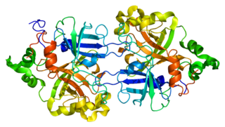
Complement component 3, often simply called C3, is a protein of the immune system that is found primarily in the blood. It plays a central role in the complement system of vertebrate animals and contributes to innate immunity. In humans it is encoded on chromosome 19 by a gene called C3.

<span class="mw-page-title-main">C5-convertase</span> Serine protease that plays key role in innate immunity.

C5 convertase is an enzyme belonging to a family of serine proteases that play key role in the innate immunity. It participates in the complement system ending with cell death.

<span class="mw-page-title-main">Thrombomodulin</span>

Thrombomodulin (TM), CD141 or BDCA-3 is an integral membrane protein expressed on the surface of endothelial cells and serves as a cofactor for thrombin. It reduces blood coagulation by converting thrombin to an anticoagulant enzyme from a procoagulant enzyme. Thrombomodulin is also expressed on human mesothelial cell, monocyte and a dendritic cell subset.

<span class="mw-page-title-main">MASP1 (protein)</span> Protein-coding gene in the species Homo sapiens

Mannan-binding lectin serine protease 1 also known as mannose-associated serine protease 1 (MASP-1) is an enzyme that in humans is encoded by the MASP1 gene.

<span class="mw-page-title-main">Factor D</span> Class of enzymes

Factor D is a protein which in humans is encoded by the CFD gene. Factor D is involved in the alternative complement pathway of the complement system where it cleaves factor B.

<span class="mw-page-title-main">Complement factor B</span> Protein-coding gene in the species Homo sapiens

Complement factor B is a protein that in humans is encoded by the CFB gene.

Complement control protein are proteins that interact with components of the complement system.

<span class="mw-page-title-main">Factor H</span> Protein-coding gene in the species Homo sapiens

Factor H (FH) is a member of the regulators of complement activation family and is a complement control protein. It is a large, soluble glycoprotein that circulates in human plasma. Its principal function is to regulate the alternative pathway of the complement system, ensuring that the complement system is directed towards pathogens or other dangerous material and does not damage host tissue. Factor H regulates complement activation on self cells and surfaces by possessing both cofactor activity for the Factor I mediated C3b cleavage, and decay accelerating activity against the alternative pathway C3-convertase, C3bBb. Factor H exerts its protective action on self cells and self surfaces but not on the surfaces of bacteria or viruses. There are however, important exceptions, such as for example the bacterial pathogen, Neisseria meningitidis. This human pathogen has evolved mechanisms to recruit human FH and down-regulate the alternative pathway. Binding of FH permits the bacteria to proliferate in the bloodstream and cause disease.

<span class="mw-page-title-main">CD46</span> Mammalian protein found in Homo sapiens

CD46 complement regulatory protein also known as CD46 and Membrane Cofactor Protein is a protein which in humans is encoded by the CD46 gene. CD46 is an inhibitory complement receptor.

<span class="mw-page-title-main">C3b</span>

C3b is the larger of two elements formed by the cleavage of complement component 3, and is considered an important part of the innate immune system. C3b is potent in opsonization: tagging pathogens, immune complexes (antigen-antibody), and apoptotic cells for phagocytosis. Additionally, C3b plays a role in forming a C3 convertase when bound to Factor B, or a C5 convertase when bound to C4b and C2b or when an additional C3b molecule binds to the C3bBb complex.

<span class="mw-page-title-main">CFHR1</span> Protein-coding gene in the species Homo sapiens

Complement factor H-related protein 1 is a protein that in humans is encoded by the CFHR1 gene.

<span class="mw-page-title-main">CFHR3</span> Protein-coding gene in the species Homo sapiens

Complement factor H-related protein 3 is a protein that in humans is encoded by the CFHR3 gene.

<span class="mw-page-title-main">CFHR4</span> Protein-coding gene in the species Homo sapiens

Complement factor H-related protein 4 is a protein that in humans is encoded by the CFHR4 gene.

<span class="mw-page-title-main">Complement component 4B</span> Protein-coding gene in the species Homo sapiens

Complement component 4B (Chido blood group) is a kind of the Complement component 4 protein that in humans is encoded by the C4B gene.

Atypical hemolytic uremic syndrome (aHUS), also known as complement-mediated hemolytic uremic syndrome, is an extremely rare, life-threatening, progressive disease that frequently has a genetic component. In most cases it can be effectively controlled by interruption of the complement cascade. Particular monoclonal antibodies, discussed later in the article, have proven efficacy in many cases.

References

  1. 1 2 3 GRCh38: Ensembl release 89: ENSG00000205403 - Ensembl, May 2017
  2. 1 2 3 GRCm38: Ensembl release 89: ENSMUSG00000058952 - Ensembl, May 2017
  3. "Human PubMed Reference:". National Center for Biotechnology Information, U.S. National Library of Medicine.
  4. "Mouse PubMed Reference:". National Center for Biotechnology Information, U.S. National Library of Medicine.
  5. Nelson RA, Jensen J, Gigli I, Tamura N (March 1966). "Methods for the separation, purification and measurement of nine components of hemolytic complement in guinea-pig serum". Immunochemistry. 3 (2): 111–35. doi:10.1016/0019-2791(66)90292-8. PMID   5960883.
  6. Lachmann PJ, Müller-Eberhard HJ (April 1968). "The demonstration in human serum of "conglutinogen-activating factor" and its effect on the third component of complement". Journal of Immunology. 100 (4): 691–8. doi:10.4049/jimmunol.100.4.691. PMID   5645214.
  7. Nilsson SC, Sim RB, Lea SM, Fremeaux-Bacchi V, Blom AM (August 2011). "Complement factor I in health and disease". Molecular Immunology (Submitted manuscript). 48 (14): 1611–20. doi:10.1016/j.molimm.2011.04.004. PMID   21529951. S2CID   37521895.
  8. Goldberger G, Bruns GA, Rits M, Edge MD, Kwiatkowski DJ (July 1987). "Human complement factor I: analysis of cDNA-derived primary structure and assignment of its gene to chromosome 4". The Journal of Biological Chemistry. 262 (21): 10065–71. doi: 10.1016/S0021-9258(18)61076-2 . PMID   2956252.
  9. Vyse TJ, Morley BJ, Bartok I, Theodoridis EL, Davies KA, Webster AD, Walport MJ (February 1996). "The molecular basis of hereditary complement factor I deficiency". The Journal of Clinical Investigation. 97 (4): 925–33. doi:10.1172/JCI118515. PMC   507137 . PMID   8613545.
  10. Julen N, Dauchel H, Lemercier C, Sim RB, Fontaine M, Ripoche J (January 1992). "In vitro biosynthesis of complement factor I by human endothelial cells". European Journal of Immunology. 22 (1): 213–7. doi:10.1002/eji.1830220131. PMID   1530917. S2CID   30130789.
  11. Whaley K (March 1980). "Biosynthesis of the complement components and the regulatory proteins of the alternative complement pathway by human peripheral blood monocytes". The Journal of Experimental Medicine. 151 (3): 501–16. doi:10.1084/jem.151.3.501. PMC   2185797 . PMID   6444659.
  12. Tsiftsoglou SA, Arnold JN, Roversi P, Crispin MD, Radcliffe C, Lea SM, Dwek RA, Rudd PM, Sim RB (November 2006). "Human complement factor I glycosylation: structural and functional characterisation of the N-linked oligosaccharides". Biochimica et Biophysica Acta (BBA) - Proteins and Proteomics. 1764 (11): 1757–66. CiteSeerX   10.1.1.712.1764 . doi:10.1016/j.bbapap.2006.09.007. PMID   17055788.
  13. "FURIN furin, paired basic amino acid cleaving enzyme [Homo sapiens (human)] - Gene - NCBI". www.ncbi.nlm.nih.gov. Retrieved 2018-03-30.
  14. "CFI complement factor I [Homo sapiens (human)] - Gene - NCBI". www.ncbi.nlm.nih.gov. Retrieved 2018-03-27.
  15. 1 2 Sanchez-Gallego JI, Groeneveld TW, Krentz S, Nilsson SC, Villoutreix BO, Blom AM (April 2012). "Analysis of binding sites on complement factor I using artificial N-linked glycosylation". The Journal of Biological Chemistry. 287 (17): 13572–83. doi: 10.1074/jbc.M111.326298 . PMC   3340171 . PMID   22393059.
  16. 1 2 Roversi P, Johnson S, Caesar JJ, McLean F, Leath KJ, Tsiftsoglou SA, Morgan BP, Harris CL, Sim RB, Lea SM (August 2011). "Structural basis for complement factor I control and its disease-associated sequence polymorphisms". Proceedings of the National Academy of Sciences of the United States of America. 108 (31): 12839–44. Bibcode:2011PNAS..10812839R. doi: 10.1073/pnas.1102167108 . PMC   3150940 . PMID   21768352.
  17. Ekdahl KN, Nilsson UR, Nilsson B (June 1990). "Inhibition of factor I by diisopropylfluorophosphate. Evidence of conformational changes in factor I induced by C3b and additional studies on the specificity of factor I". Journal of Immunology. 144 (11): 4269–74. doi:10.4049/jimmunol.144.11.4269. PMID   2140392.
  18. Wang Q, Zhao HS, Li L (2016-02-18). "Association between complement factor I gene polymorphisms and the risk of age-related macular degeneration: a Meta-analysis of literature". International Journal of Ophthalmology. 9 (2): 298–305. doi:10.18240/ijo.2016.02.23. PMC   4761747 . PMID   26949655.
  19. Tan PL, Garrett ME, Willer JR, Campochiaro PA, Campochiaro B, Zack DJ, Ashley-Koch AE, Katsanis N (March 2017). "Systematic Functional Testing of Rare Variants: Contributions of CFI to Age-Related Macular Degeneration". Investigative Ophthalmology & Visual Science. 58 (3): 1570–1576. doi:10.1167/iovs.16-20867. PMC   6022411 . PMID   28282489.
  20. 1 2 Kavanagh D, Goodship TH, Richards A (November 2013). "Atypical hemolytic uremic syndrome". Seminars in Nephrology. 33 (6): 508–30. doi:10.1016/j.semnephrol.2013.08.003. PMC   3863953 . PMID   24161037.

Further reading