Factor D

Last updated
complement factor D (adipsin)
Factor D.jpg
Factor D in Homo sapiens [1] [2]
Identifiers
SymbolCFD
Alt. symbolsDF, PFD
NCBI gene 1675
HGNC 2771
OMIM 134350
RefSeq NM_001928
UniProt P00746
Other data
Locus Chr. 19 p13.3
Search for
Structures Swiss-model
Domains InterPro

Factor D (EC 3.4.21.46, C3 proactivator convertase, properdin factor D esterase, factor D (complement), complement factor D, CFD, adipsin) is a protein which in humans is encoded by the CFD gene. [3] Factor D is involved in the alternative complement pathway of the complement system where it cleaves factor B.

Contents

Function

The protein encoded by this gene is a member of the trypsin family of serine proteases secreted by adipocytes into the bloodstream. The encoded protein is a component of the alternative complement pathway best known for its role in humoral suppression of infectious agents. Finally, the encoded protein has a high level of expression in fat, suggesting a role for adipose tissue in immune system biology. [3]

Alternative pathway. ( 4. Is factor D cleaving B to Bb and Ba) Droga alternatywna.png
Alternative pathway. ( 4. Is factor D cleaving B to Bb and Ba)

Factor D is a serine protease that stimulates glucose transport for triglyceride accumulation in fats cells and inhibits lipolysis. [4]

Clinical significance

The level of Factor D is decreased [5] in obese patients. This reduction may be due to high activity or resistance but exact cause is not fully known.

Structure

All members of the chymotrypsin family of serine proteases have very similar structures. In all cases, including factor D, there are two antiparallel β-barrel domains with each barrel containing six β-strands with the same typology in all enzymes. The major difference in backbone structure between Factor D and the other serine proteases of the chymotrypsin family is in the surface loops connecting the secondary structural elements. Factor D displays different conformations of major catalytic and substrate-binding residues typically found in the chrotrypsin family. These features suggest the catalytic activity of factor D is prohibited unless conformational changes are induced by a realignment. [6]

Mechanism of Action

Factor D is a serine protease present in blood and tissue in an active sequence but self-inhibited conformation. The only known natural substrate of Factor D is Factor B, and cleavage of the Arg234-Lys235 scissile bond in Factor B results in two Factor B fragments, Ba and Bb. Before cleavage of the scissile bond in Factor B can occur, Factor B must first bind with C3b before to form the C3bB complex. [7] It is proposed that this conformational change of Factor B in the C3bB complex allows Factor B to fit into the binding site of Factor D.

The catalytic triad of Factor D is composed of Asp102, His57 and Ser195. Other key components of Factor D are an Asp189-Arg218 salt bridge that stabilizes a self-inhibitory loop (amino acid residues 212 to 218) and His57 side chain in the non-canonical conformation. [8] [9] In its inhibited form, the self-inhibitory loop prevents access of Factor B to Factor D. When the self-inhibited conformation of Factor D is approached by the C3bB complex, C3bB displaces the salt bridge in Factor D and results in a new salt bridge between the Arg234 of Factor B and Asp189 of Factor D. [10] [11] The displacement of the Factor D salt bridge results in a realignment of the self-inhibitory loop and a rotation of the active site histidine side chain, creating the canonical form of Factor D. Cleavage of the scissile bond in Factor B then ensues, releasing fragment Ba and forming C3bBb, the alternative pathway C3-convertase. [12]

The non-canonical conformation of Factor D is inhibited by the self-inhibitory loop (blue). The Asp-Arg salt bridge (purple and orange side chains, respectively) stabilizes the self-inhibitory loop. The catalytic triad is shown in green. Factor D Non-Canonical.png
The non-canonical conformation of Factor D is inhibited by the self-inhibitory loop (blue). The Asp-Arg salt bridge (purple and orange side chains, respectively) stabilizes the self-inhibitory loop. The catalytic triad is shown in green.
The canonical conformation of Factor D is not self-inhibited. The Asp-Arg salt bridge (purple and orange side chains, respectively) has been displaced resulting in a shift in the self inhibitory loop (blue). The catalytic triad is shown in green. Factor D Canonical Configuration.png
The canonical conformation of Factor D is not self-inhibited. The Asp-Arg salt bridge (purple and orange side chains, respectively) has been displaced resulting in a shift in the self inhibitory loop (blue). The catalytic triad is shown in green.

Regulation

Factor D is synthesized by the liver and adipocytes with the latter being the major source. The pro-form of Factor D that is secreted is cleaved by MASP-3 to form the active sequence that circulates in the body. [15] Factor D maintains an extremely high substrate specificity, and as a result has no known natural inhibitors in the body. [16] However, most of Factor D remains in the self-inhibited form that limits substrate access to the catalytic site. Factor D has a molecular weight of 23.5 kD and is present at a concentration of 1.8 mg/L of blood in healthy humans. The synthesis rate of Factor is approximately 1.33 mg/kg/day, and most of Factor D is eliminated through the kidney after catabolism in proximal tubules after re-absorption. The net effect is a high fractional metabolic rate of 60% per hour. [17] In patients with normal kidney function, no Factor D was detectable in urine. However, in patients with renal disease, Factor D was found at elevated levels. The alternative pathway is capable of operating even at low levels of Factor D, and deficiencies in levels of Factor D are rare. [18] [19]

Role in Diseases

A point mutation resulting in the replacement of a serine codon (Ser42 in the unprocessed methionine form of Factor D) with a stop codon (TAG) in the Factor D gene on chromosome 19 has been documented as a cause of Factor D deficiency. [20] Deficiency in Factor D may cause an increased susceptibility to bacterial infections, specifically Neisseria infections. The mode of inheritance of Factor D deficiency is autosomal recessive, and individuals with a mutation on only one allele may not experience the same susceptibility to reoccurring infections. In a patient with reoccurring infections, complete improvement in the condition was obtained by introducing purified Factor D. [21]

Diseases with excessive complement activation include paroxysmal nocturnal hemoglobinuria (PNH), and inhibitors of Factor D may have utility in the treatment of PNH. Small molecule inhibitors of Factor D are under development for the treatment of PNH, and one small molecule inhibitor, ACH-4471, has shown promise in a Phase 2 clinical trial for Factor D inhibition when combined with eculizumab. Patients treated with Factor D inhibitors must be immunized against infections in order to avoid reoccurring infections as in patients with Factor D deficiency. [22] [23]

Related Research Articles

<span class="mw-page-title-main">Protease</span> Enzyme that cleaves other proteins into smaller peptides

A protease is an enzyme that catalyzes proteolysis, breaking down proteins into smaller polypeptides or single amino acids, and spurring the formation of new protein products. They do this by cleaving the peptide bonds within proteins by hydrolysis, a reaction where water breaks bonds. Proteases are involved in many biological functions, including digestion of ingested proteins, protein catabolism, and cell signaling.

<span class="mw-page-title-main">Antithrombin</span> Mammalian protein found in Homo sapiens

Antithrombin (AT) is a small glycoprotein that inactivates several enzymes of the coagulation system. It is a 464-amino-acid protein produced by the liver. It contains three disulfide bonds and a total of four possible glycosylation sites. α-Antithrombin is the dominant form of antithrombin found in blood plasma and has an oligosaccharide occupying each of its four glycosylation sites. A single glycosylation site remains consistently un-occupied in the minor form of antithrombin, β-antithrombin. Its activity is increased manyfold by the anticoagulant drug heparin, which enhances the binding of antithrombin to factor IIa (thrombin) and factor Xa.

<span class="mw-page-title-main">Complement system</span> Part of the immune system that enhances the ability of antibodies and phagocytic cells

The complement system, also known as complement cascade, is a part of the immune system that enhances (complements) the ability of antibodies and phagocytic cells to clear microbes and damaged cells from an organism, promote inflammation, and attack the pathogen's cell membrane. It is part of the innate immune system, which is not adaptable and does not change during an individual's lifetime. The complement system can, however, be recruited and brought into action by antibodies generated by the adaptive immune system.

<span class="mw-page-title-main">Serpin</span> Superfamily of proteins with similar structures and diverse functions

Serpins are a superfamily of proteins with similar structures that were first identified for their protease inhibition activity and are found in all kingdoms of life. The acronym serpin was originally coined because the first serpins to be identified act on chymotrypsin-like serine proteases. They are notable for their unusual mechanism of action, in which they irreversibly inhibit their target protease by undergoing a large conformational change to disrupt the target's active site. This contrasts with the more common competitive mechanism for protease inhibitors that bind to and block access to the protease active site.

<span class="mw-page-title-main">Serine protease</span> Class of enzymes

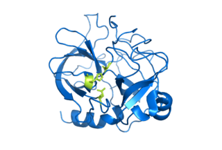
Serine proteases are enzymes that cleave peptide bonds in proteins. Serine serves as the nucleophilic amino acid at the (enzyme's) active site. They are found ubiquitously in both eukaryotes and prokaryotes. Serine proteases fall into two broad categories based on their structure: chymotrypsin-like (trypsin-like) or subtilisin-like.

<span class="mw-page-title-main">Classical complement pathway</span> Aspect of the immune system

The classical complement pathway is one of three pathways which activate the complement system, which is part of the immune system. The classical complement pathway is initiated by antigen-antibody complexes with the antibody isotypes IgG and IgM.

<span class="mw-page-title-main">C3-convertase</span>

C3 convertase belongs to family of serine proteases and is necessary in innate immunity as a part of the complement system which eventuate in opsonisation of particles, release of inflammatory peptides, C5 convertase formation and cell lysis.

<span class="mw-page-title-main">Plasminogen activator</span> Type of protein

Plasminogen activators are serine proteases that catalyze the activation of plasmin via proteolytic cleavage of its zymogen form plasminogen. Plasmin is an important factor in fibrinolysis, the breakdown of fibrin polymers formed during blood clotting. There are two main plasminogen activators: urokinase (uPA) and tissue plasminogen activator (tPA). Tissue plasminogen activators are used to treat medical conditions related to blood clotting including embolic or thrombotic stroke, myocardial infarction, and pulmonary embolism.

<span class="mw-page-title-main">Complement component 3</span> Protein-coding gene in the species Homo sapiens

Complement component 3, often simply called C3, is a protein of the immune system that is found primarily in the blood. It plays a central role in the complement system of vertebrate animals and contributes to innate immunity. In humans it is encoded on chromosome 19 by a gene called C3.

<span class="mw-page-title-main">C5-convertase</span> Serine protease that plays key role in innate immunity.

C5 convertase is an enzyme belonging to a family of serine proteases that play key role in the innate immunity. It participates in the complement system ending with cell death.

<span class="mw-page-title-main">Lectin pathway</span>

The lectin pathway or lectin complement pathway is a type of cascade reaction in the complement system, similar in structure to the classical complement pathway, in that, after activation, it proceeds through the action of C4 and C2 to produce activated complement proteins further down the cascade. In contrast to the classical complement pathway, the lectin pathway does not recognize an antibody bound to its target. The lectin pathway starts with mannose-binding lectin (MBL) or ficolin binding to certain sugars.

<span class="mw-page-title-main">Complement component 2</span> Protein-coding gene in the species Homo sapiens

Complement C2 is a protein that in humans is encoded by the C2 gene. The protein encoded by this gene is part of the classical pathway of the complement system, acting as a multi-domain serine protease. Deficiency of C2 has been associated with certain autoimmune diseases.

<span class="mw-page-title-main">Complement component 1s</span> Protein-coding gene in the species Homo sapiens

Complement component 1s is a protein involved in the complement system. C1s is part of the C1 complex. In humans, it is encoded by the C1S gene.

<span class="mw-page-title-main">MASP1 (protein)</span> Protein-coding gene in the species Homo sapiens

Mannan-binding lectin serine protease 1 also known as mannose-associated serine protease 1 (MASP-1) is an enzyme that in humans is encoded by the MASP1 gene.

<span class="mw-page-title-main">MASP2 (protein)</span> Protein-coding gene in the species Homo sapiens

Mannan-binding lectin serine protease 2 also known as mannose-binding protein-associated serine protease 2 (MASP-2) is an enzyme that in humans is encoded by the MASP2 gene.

<span class="mw-page-title-main">Complement factor B</span> Protein-coding gene in the species Homo sapiens

Complement factor B is a protein that in humans is encoded by the CFB gene.

<span class="mw-page-title-main">Complement factor I</span> Protein

Complement factor I, also known as C3b/C4b inactivator, is a protein that in humans is encoded by the CFI gene. Complement factor I is a protein of the complement system, first isolated in 1966 in guinea pig serum, that regulates complement activation by cleaving cell-bound or fluid phase C3b and C4b. It is a soluble glycoprotein that circulates in human blood at an average concentration of 35 μg/mL.

<span class="mw-page-title-main">SPINT1</span> Protein-coding gene in the species Homo sapiens

Kunitz-type protease inhibitor 1 is an enzyme that in humans is encoded by the SPINT1 gene.

<span class="mw-page-title-main">Glutamyl endopeptidase GluV8</span>

Glutamyl endopeptidase is an extracellular bacterial serine protease of the glutamyl endopeptidase I family that was initially isolated from the Staphylococcus aureus strain V8. The protease is, hence, commonly referred to as "V8 protease", or alternatively SspA from its corresponding gene.

<span class="mw-page-title-main">Aureolysin</span>

Aureolysin is an extracellular metalloprotease expressed by Staphylococcus aureus. This protease is a major contributor to the bacterium's virulence, or ability to cause disease, by cleaving host factors of the innate immune system as well as regulating S. aureus secreted toxins and cell wall proteins. To catalyze its enzymatic activities, aureolysin requires zinc and calcium which it obtains from the extracellular environment within the host.

References

  1. PDB: 1HFD
  2. Narayana SV, Carson M, el-Kabbani O, Kilpatrick JM, Moore D, Chen X, Bugg CE, Volanakis JE, DeLucas LJ (1994). "Structure of human factor D. A complement system protein at 2.0 A resolution". Journal of Molecular Biology. 235 (2): 695–708. doi:10.1006/jmbi.1994.1021. PMID   8289289.
  3. 1 2 EntrezGene 1675
  4. Ronti T, Lupattelli G, Mannarino E (2006). "The endocrine function of adipose tissue: an update". Clinical Endocrinology. 64 (4): 355–65. doi:10.1111/j.1365-2265.2006.02474.x. PMID   16584505. S2CID   12455240.
  5. Flier JS, Cook KS, Usher P, Spiegelman BM (1987). "Severely impaired adipsin expression in genetic and acquired obesity". Science. 237 (4813): 405–8. Bibcode:1987Sci...237..405F. doi:10.1126/science.3299706. PMID   3299706.
  6. Volanakis JE, Narayana SV (1996). "Complement factor D, a novel serine protease". Protein Science. 5 (4): 553–64. doi:10.1002/pro.5560050401. PMC   2143395 . PMID   8845746.
  7. Lesavre, PH; Müller-Eberhard, HJ (1 December 1978). "Mechanism of action of factor D of the alternative complement pathway". The Journal of Experimental Medicine. 148 (6): 1498–509. doi:10.1084/jem.148.6.1498. PMC   2185104 . PMID   82604.
  8. Jing, H; Babu, YS; Moore, D; Kilpatrick, JM; Liu, XY; Volanakis, JE; Narayana, SV (9 October 1998). "Structures of native and complexed complement factor D: implications of the atypical His57 conformation and self-inhibitory loop in the regulation of specific serine protease activity". Journal of Molecular Biology. 282 (5): 1061–81. doi:10.1006/jmbi.1998.2089. PMID   9753554.
  9. Jing, H; Macon, KJ; Moore, D; DeLucas, LJ; Volanakis, JE; Narayana, SV (15 February 1999). "Structural basis of profactor D activation: from a highly flexible zymogen to a novel self-inhibited serine protease, complement factor D." The EMBO Journal. 18 (4): 804–14. doi:10.1093/emboj/18.4.804. PMC   1171173 . PMID   10022823.
  10. Karki, RG; Powers, J; Mainolfi, N; Anderson, K; Belanger, DB; Liu, D; Ji, N; Jendza, K; Gelin, CF; Mac Sweeney, A; Solovay, C; Delgado, O; Crowley, M; Liao, SM; Argikar, UA; Flohr, S; La Bonte, LR; Lorthiois, EL; Vulpetti, A; Brown, A; Long, D; Prentiss, M; Gradoux, N; de Erkenez, A; Cumin, F; Adams, C; Jaffee, B; Mogi, M (9 May 2019). "Design, Synthesis, and Preclinical Characterization of Selective Factor D Inhibitors Targeting the Alternative Complement Pathway". Journal of Medicinal Chemistry. 62 (9): 4656–4668. doi:10.1021/acs.jmedchem.9b00271. PMID   30995036. S2CID   122356241.
  11. Forneris, F; Ricklin, D; Wu, J; Tzekou, A; Wallace, RS; Lambris, JD; Gros, P (24 December 2010). "Structures of C3b in complex with factors B and D give insight into complement convertase formation". Science. 330 (6012): 1816–20. Bibcode:2010Sci...330.1816F. doi:10.1126/science.1195821. PMC   3087196 . PMID   21205667.
  12. Vulpetti, A; Randl, S; Rüdisser, S; Ostermann, N; Erbel, P; Mac Sweeney, A; Zoller, T; Salem, B; Gerhartz, B; Cumin, F; Hommel, U; Dalvit, C; Lorthiois, E; Maibaum, J (9 March 2017). "Structure-Based Library Design and Fragment Screening for the Identification of Reversible Complement Factor D Protease Inhibitors". Journal of Medicinal Chemistry. 60 (5): 1946–1958. doi:10.1021/acs.jmedchem.6b01684. PMID   28157311.
  13. Maibaum, J; Liao, SM; Vulpetti, A; Ostermann, N; Randl, S; Rüdisser, S; Lorthiois, E; Erbel, P; Kinzel, B; Kolb, FA; Barbieri, S; Wagner, J; Durand, C; Fettis, K; Dussauge, S; Hughes, N; Delgado, O; Hommel, U; Gould, T; Mac Sweeney, A; Gerhartz, B; Cumin, F; Flohr, S; Schubart, A; Jaffee, B; Harrison, R; Risitano, AM; Eder, J; Anderson, K (December 2016). "Small-molecule factor D inhibitors targeting the alternative complement pathway". Nature Chemical Biology. 12 (12): 1105–1110. doi:10.1038/nchembio.2208. PMID   27775713.
  14. Vulpetti, A; Ostermann, N; Randl, S; Yoon, T; Mac Sweeney, A; Cumin, F; Lorthiois, E; Rüdisser, S; Erbel, P; Maibaum, J (10 May 2018). "Discovery and Design of First Benzylamine-Based Ligands Binding to an Unlocked Conformation of the Complement Factor D." ACS Medicinal Chemistry Letters. 9 (5): 490–495. doi:10.1021/acsmedchemlett.8b00104. PMC   5949727 . PMID   29795765.
  15. Hayashi, M; Machida, T; Ishida, Y; Ogata, Y; Omori, T; Takasumi, M; Endo, Y; Suzuki, T; Sekimata, M; Homma, Y; Ikawa, M; Ohira, H; Fujita, T; Sekine, H (15 September 2019). "Cutting Edge: Role of MASP-3 in the Physiological Activation of Factor D of the Alternative Complement Pathway". Journal of Immunology. 203 (6): 1411–1416. doi: 10.4049/jimmunol.1900605 . PMID   31399515. S2CID   199518699.
  16. Lorthiois, E; Anderson, K; Vulpetti, A; Rogel, O; Cumin, F; Ostermann, N; Steinbacher, S; Mac Sweeney, A; Delgado, O; Liao, SM; Randl, S; Rüdisser, S; Dussauge, S; Fettis, K; Kieffer, L; de Erkenez, A; Yang, L; Hartwieg, C; Argikar, UA; La Bonte, LR; Newton, R; Kansara, V; Flohr, S; Hommel, U; Jaffee, B; Maibaum, J (13 July 2017). "Discovery of Highly Potent and Selective Small-Molecule Reversible Factor D Inhibitors Demonstrating Alternative Complement Pathway Inhibition in Vivo". Journal of Medicinal Chemistry. 60 (13): 5717–5735. doi:10.1021/acs.jmedchem.7b00425. PMID   28621538.
  17. Dobó, J; Kocsis, A; Gál, P (2018). "Be on Target: Strategies of Targeting Alternative and Lectin Pathway Components in Complement-Mediated Diseases". Frontiers in Immunology. 9: 1851. doi: 10.3389/fimmu.2018.01851 . PMC   6092519 . PMID   30135690.
  18. Volanakis, JE; Barnum, SR; Giddens, M; Galla, JH (14 February 1985). "Renal filtration and catabolism of complement protein D.". The New England Journal of Medicine. 312 (7): 395–9. doi:10.1056/NEJM198502143120702. PMID   3844050.
  19. Pascual, M; Steiger, G; Estreicher, J; Macon, K; Volanakis, JE; Schifferli, JA (October 1988). "Metabolism of complement factor D in renal failure". Kidney International. 34 (4): 529–36. doi: 10.1038/ki.1988.214 . PMID   3199673.
  20. Biesma, DH; Hannema, AJ; van Velzen-Blad, H; Mulder, L; van Zwieten, R; Kluijt, I; Roos, D (July 2001). "A family with complement factor D deficiency". The Journal of Clinical Investigation. 108 (2): 233–40. doi:10.1172/JCI12023. PMC   203023 . PMID   11457876.
  21. Hiemstra, PS; Langeler, E; Compier, B; Keepers, Y; Leijh, PC; van den Barselaar, MT; Overbosch, D; Daha, MR (December 1989). "Complete and partial deficiencies of complement factor D in a Dutch family". The Journal of Clinical Investigation. 84 (6): 1957–61. doi:10.1172/JCI114384. PMC   304077 . PMID   2687330.
  22. Yuan, X; Gavriilaki, E; Thanassi, JA; Yang, G; Baines, AC; Podos, SD; Huang, Y; Huang, M; Brodsky, RA (March 2017). "Small-molecule factor D inhibitors selectively block the alternative pathway of complement in paroxysmal nocturnal hemoglobinuria and atypical hemolytic uremic syndrome". Haematologica. 102 (3): 466–475. doi:10.3324/haematol.2016.153312. PMC   5394948 . PMID   27810992.
  23. Risitano, AM (January 2014). "Anti-Complement Treatment in Paroxysmal Nocturnal Hemoglobinuria: Where we Stand and Where we are Going". Translational Medicine @ UniSa. 8: 43–52. PMC   4000462 . PMID   24778997.

This article incorporates text from the United States National Library of Medicine, which is in the public domain.