Lemierre's syndrome

Last updated
Lemierre's syndrome
Other namesSeptic phlebitis of the internal jugular vein, postanginal sepsis secondary to oropharyngeal infection, postanginal shock including sepsis, Lemierre's disease, human necrobacillosis
Fusobacterium novum 01.jpg
Fusobacterium necrophorum, the most common cause of Lemierre's syndrome
Specialty Infectious diseases, veterinary medicine   OOjs UI icon edit-ltr-progressive.svg
Symptoms Early:Fever, sore throat, fatigue Later:Variable, Sepsis, vomiting, muscle pain, meningitis, hepatosplenomegaly
Complications Septic shock, kidney failure, liver failure, cerebral edema, organ failure [1]

Lemierre's syndrome is infectious thrombophlebitis of the internal jugular vein. [2] It most often develops as a complication of a bacterial sore throat infection in young, otherwise healthy adults. The thrombophlebitis is a serious condition and may lead to further systemic complications such as bacteria in the blood or septic emboli.

Contents

Lemierre's syndrome occurs most often when a bacterial (e.g., Fusobacterium necrophorum ) throat infection progresses to the formation of a peritonsillar abscess. Deep in the abscess, anaerobic bacteria can flourish. When the abscess wall ruptures internally, the drainage carrying bacteria seeps through the soft tissue and infects the nearby structures. Spread of infection to the nearby internal jugular vein provides a gateway for the spread of bacteria through the bloodstream. The inflammation surrounding the vein and compression of the vein may lead to blood clot formation. Pieces of the potentially infected clot can break off and travel through the right heart into the lungs as emboli, blocking branches of the pulmonary artery that carry blood with little oxygen from the right side of the heart to the lungs.[ citation needed ]

Sepsis following a throat infection was first described by Hugo Schottmüller in 1918. [3] In 1936, André Lemierre published a series of 20 cases where throat infections were followed by identified anaerobic sepsis, of whom 18 died. [4]

Signs and symptoms

The signs and symptoms of Lemierre's syndrome vary, but usually start with a sore throat, fever, and general body weakness. These are followed by extreme lethargy, spiked fevers, rigors, swollen cervical lymph nodes, and a swollen, tender or painful neck. Often there is abdominal pain, diarrhea, nausea and vomiting during this phase. These signs and symptoms usually occur several days to two weeks after the initial symptoms. Symptoms of pulmonary involvement can be shortness of breath, cough and painful breathing (pleuritic chest pain). Rarely, blood is coughed up. Painful or inflamed joints can occur when the joints are involved.[ citation needed ]

Septic shock can also arise. This presents with low blood pressure, increased heart rate, decreased urine output and an increased rate of breathing. Some cases will also present with meningitis, which will typically manifest as neck stiffness, headache and sensitivity of the eyes to light.[ citation needed ] Liver enlargement and spleen enlargement can be found, but are not always associated with liver or spleen abscesses. [5] [6] Other signs and symptoms that may occur:[ citation needed ]

Cause

The bacteria causing the thrombophlebitis are anaerobic bacteria that are typically normal components of the microorganisms that inhabit the mouth and throat. Species of Fusobacterium , specifically Fusobacterium necrophorum , are most commonly the causative bacteria, but various bacteria have been implicated. One 1989 study found that 81% of Lemierres's syndrome had been infected with Fusobacterium necrophorum, while 11% were caused by other Fusobacterium species. [7] MRSA might also be an issue in Lemierre infections. [8] Rarely Lemierre's syndrome is caused by other (usually Gram-negative) bacteria, which include Bacteroides fragilis and Bacteroides melaninogenicus , Peptostreptococcus spp., Streptococcus microaerophile , Staphylococcus aureus , Streptococcus pyogenes , and Eikenella corrodens . [7] [9]

Pathophysiology

Lemierre's syndrome begins with an infection of the head and neck region, with most primary sources of infection in the palatine tonsils and peritonsillar tissue. [10] Usually this infection is a pharyngitis (which occurred in 87.1% of patients as reported by a literature review [6] ), and can be preceded by infectious mononucleosis as reported in several cases. [10] It can also be initiated by infections of the ear, mastoid bone, sinuses, or saliva glands.[ citation needed ]

During the primary infection, F. necrophorum colonizes the infection site and the infection spreads to the parapharyngeal space. The bacteria then invade the peritonsillar blood vessels where they can spread to the internal jugular vein. [5] In this vein, the bacteria cause the formation of a thrombus containing these bacteria. Furthermore, the internal jugular vein becomes inflamed. This septic thrombophlebitis can give rise to septic microemboli [11] that disseminate to other parts of the body where they can form abscesses and septic infarctions. The first capillaries that the emboli encounter where they can nestle themselves are the pulmonary capillaries. As a consequence, the most frequently involved site of septic metastases are the lungs, followed by the joints (knee, hip, sternoclavicular joint, shoulder and elbow [12] ). In the lungs, the bacteria cause abscesses, nodulary and cavitary lesions. Pleural effusion is often present. [6] Other sites involved in septic metastasis and abscess formation are the muscles and soft tissues, liver, spleen, kidneys and nervous system (intracranial abscesses, meningitis). [5]

Production of bacterial toxins such as lipopolysaccharide leads to secretion of cytokines by white blood cells which then both lead to symptoms of sepsis. F. necrophorum produces hemagglutinin which causes platelet aggregation that can lead to diffuse intravascular coagulation and thrombocytopenia. [13] [14]

Diagnosis

Diagnosis and the imaging (and laboratory) studies to be ordered largely depend on the patient history, signs and symptoms. If a persistent sore throat with signs of sepsis are found, physicians are cautioned to screen for Lemierre's syndrome.[ citation needed ]

Laboratory investigations reveal signs of a bacterial infection with elevated C-reactive protein, erythrocyte sedimentation rate and white blood cells (notably neutrophils). Platelet count can be low or high. Liver and kidney function tests are often abnormal.[ citation needed ]

Thrombosis of the internal jugular vein can be displayed with sonography. Thrombi that have developed recently have low echogenicity or echogenicity similar to the flowing blood, and in such cases pressure with the ultrasound probe show a non-compressible jugular vein - a sure sign of thrombosis. Also color or power Doppler ultrasound identify a low echogenicity blood clot. A CT scan or an MRI scan is more sensitive in displaying the thrombus of the intra-thoracic retrosternal veins, but are rarely needed.[ citation needed ]

Chest X-ray and chest CT may show pleural effusion, nodules, infiltrates, abscesses and cavitations.[ citation needed ]

Bacterial cultures taken from the blood, joint aspirates or other sites can identify the causative agent of the disease.[ citation needed ]

Other illnesses that can be included in the differential diagnosis are:[ citation needed ]

Treatment

Lemierre's syndrome is primarily treated with antibiotics given intravenously. Fusobacterium necrophorum is generally highly susceptible to beta-lactam antibiotics, metronidazole, clindamycin and third generation cephalosporins while the other fusobacteria have varying degrees of resistance to beta-lactams and clindamycin. [14] Additionally, there may exist a co-infection by another bacterium. For these reasons is often advised not to use monotherapy in treating Lemierre's syndrome. Penicillin and penicillin-derived antibiotics can thus be combined with a beta-lactamase inhibitor such as clavulanic acid or with metronidazole. [5] [9] Clindamycin can be given as monotherapy.[ citation needed ]

If antibiotic therapy is unsuccessful, additional treatments include draining of any abscesses and ligation of the internal jugular vein where the antibiotic cannot penetrate. [6] [9] [15] There is no evidence to opt for or against the use of anticoagulation therapy. The low incidence of Lemierre's syndrome has not made it possible to set up clinical trials to study the disease. [9]

Prognosis

The mortality rate was 90% prior to antibiotic therapy. In the contemporary era, a mortality of 4% has been estimated. [16] Since this disease is not well known and often remains undiagnosed, mortality might be much higher. Approximately 10% of those with the condition experience clinical sequelae, including cranial nerve palsy and orthopaedic limitations. [16]

Epidemiology

Lemierre's syndrome is currently rare, but was more common in the early 20th century before the discovery of penicillin. The reduced use of antibiotics for sore throats may have increased the risk of this disease, with 19 cases in 1997 and 34 cases in 1999 reported in the UK. [17] The estimated incidence rate is 0.8 to 3.6 cases per million in the general population, but is higher in healthy young adults. The number of cases reported is increasing; however, because of its rarity, physicians may be unaware of its existence, possibly leading to underdiagnosis. [18]

History

Sepsis following from a throat infection was described by Hugo Schottmüller in 1918. [3] In 1936, André Lemierre published a series of 20 cases where throat infections were followed by identified anaerobic sepsis, of whom 18 patients died. [4]

Related Research Articles

<span class="mw-page-title-main">Brain abscess</span> Medical condition

Brain abscess is an abscess within the brain tissue caused by inflammation and collection of infected material coming from local or remote infectious sources. The infection may also be introduced through a skull fracture following a head trauma or surgical procedures. Brain abscess is usually associated with congenital heart disease in young children. It may occur at any age but is most frequent in the third decade of life.

<span class="mw-page-title-main">Danger triangle of the face</span> Area of the face with blood supply that connects indirectly to the brain

The danger triangle of the face consists of the area from the corners of the mouth to the bridge of the nose, including the nose and maxilla. Due to the special nature of the blood supply to the human nose and surrounding area, it is possible, albeit extremely unlikely, for retrograde infection from the nasal area to spread to the brain, causing cavernous sinus thrombosis, meningitis, or brain abscess.

<i>Fusobacterium</i> Genus of bacteria

Fusobacterium is a genus of obligate anaerobic, Gram-negative, non-sporeforming bacteria belonging to Gracilicutes. Individual cells are slender, rod-shaped bacilli with pointed ends. Fusobacterium was discovered in 1900 by Courmont and Cade and is common in the flora of humans.

<span class="mw-page-title-main">Pharyngitis</span> Inflammation of the back of the throat

Pharyngitis is inflammation of the back of the throat, known as the pharynx. It typically results in a sore throat and fever. Other symptoms may include a runny nose, cough, headache, difficulty swallowing, swollen lymph nodes, and a hoarse voice. Symptoms usually last 3–5 days, but can be longer depending on cause. Complications can include sinusitis and acute otitis media. Pharyngitis is a type of upper respiratory tract infection.

Bloodstream infections (BSIs), septicemia which include bacteremias when the infections are bacterial and fungemias when the infections are fungal, are infections present in the blood. Blood is normally a sterile environment, so the detection of microbes in the blood is always abnormal. A bloodstream infection is different from sepsis, which is the host response to bacteria.

<span class="mw-page-title-main">Septic shock</span> Dangerously low blood pressure due to damage from an organ infection

Septic shock is a potentially fatal medical condition that occurs when sepsis, which is organ injury or damage in response to infection, leads to dangerously low blood pressure and abnormalities in cellular metabolism. The Third International Consensus Definitions for Sepsis and Septic Shock (Sepsis-3) defines septic shock as a subset of sepsis in which particularly profound circulatory, cellular, and metabolic abnormalities are associated with a greater risk of mortality than with sepsis alone. Patients with septic shock can be clinically identified by requiring a vasopressor to maintain a mean arterial pressure of 65 mm Hg or greater and having serum lactate level greater than 2 mmol/L (>18 mg/dL) in the absence of hypovolemia. This combination is associated with hospital mortality rates greater than 40%.

<span class="mw-page-title-main">Cellulitis</span> Bacterial infection of the inner layers of the skin called the dermis

Cellulitis is usually a bacterial infection involving the inner layers of the skin. It specifically affects the dermis and subcutaneous fat. Signs and symptoms include an area of redness which increases in size over a few days. The borders of the area of redness are generally not sharp and the skin may be swollen. While the redness often turns white when pressure is applied, this is not always the case. The area of infection is usually painful. Lymphatic vessels may occasionally be involved, and the person may have a fever and feel tired.

<span class="mw-page-title-main">Tonsillitis</span> Inflammation of the tonsils

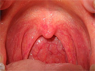
Tonsillitis is inflammation of the tonsils in the upper part of the throat. It can be acute or chronic. Acute tonsillitis typically has a rapid onset. Symptoms may include sore throat, fever, enlargement of the tonsils, trouble swallowing, and enlarged lymph nodes around the neck. Complications include peritonsillar abscess (Quinsy).

<span class="mw-page-title-main">Peritonsillar abscess</span> Pus behind the tonsil due to an infection

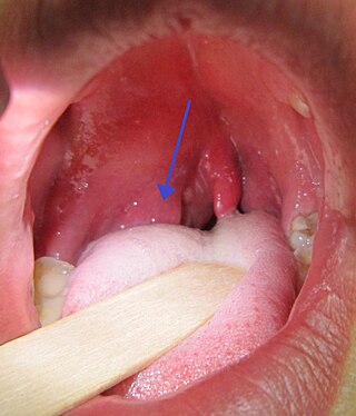
Peritonsillar abscess (PTA), also known as quinsy, is an accumulation of pus due to an infection behind the tonsil. Symptoms include fever, throat pain, trouble opening the mouth, and a change to the voice. Pain is usually worse on one side. Complications may include blockage of the airway or aspiration pneumonitis.

<span class="mw-page-title-main">Mastoiditis</span> Middle ear disease

Mastoiditis is the result of an infection that extends to the air cells of the skull behind the ear. Specifically, it is an inflammation of the mucosal lining of the mastoid antrum and mastoid air cell system inside the mastoid process. The mastoid process is the portion of the temporal bone of the skull that is behind the ear. The mastoid process contains open, air-containing spaces. Mastoiditis is usually caused by untreated acute otitis media and used to be a leading cause of child mortality. With the development of antibiotics, however, mastoiditis has become quite rare in developed countries where surgical treatment is now much less frequent and more conservative, unlike former times.

<span class="mw-page-title-main">Lung abscess</span> Medical condition

Lung abscess is a type of liquefactive necrosis of the lung tissue and formation of cavities containing necrotic debris or fluid caused by microbial infection.

<span class="mw-page-title-main">Lymphangitis</span> Medical condition

Lymphangitis is an inflammation or an infection of the lymphatic channels that occurs as a result of infection at a site distal to the channel. The most common cause of lymphangitis in humans is Streptococcus pyogenes, hemolytic streptococci, and in some cases, mononucleosis, cytomegalovirus, tuberculosis, syphilis, and the fungus Sporothrix schenckii. Lymphangitis is sometimes mistakenly called "blood poisoning". In reality, "blood poisoning" is synonymous with sepsis.

<span class="mw-page-title-main">Cavernous sinus thrombosis</span> Medical condition

Cavernous sinus thrombosis (CST) is the formation of a blood clot within the cavernous sinus, a cavity at the base of the brain which drains deoxygenated blood from the brain back to the heart. This is a rare disorder and can be of two types–septic cavernous thrombosis and aseptic cavernous thrombosis. The most common form is septic cavernous sinus thrombosis. The cause is usually from a spreading infection in the nose, sinuses, ears, or teeth. Staphylococcus aureus and Streptococcus are often the associated bacteria.

Fusobacterium necrophorum is a species of bacteria responsible for Lemierre's syndrome. It has also been known to cause sinusitis, mastoiditis, and odontogenic infections.

A septic embolism is a type of embolism that is infected with bacteria, resulting in the formation of pus. These may become dangerous if dislodged from their original location. Like other emboli, a septic embolism may be fatal.

Septic abortion describes any type of abortion, due to an upper genital tract bacterial infection including the inflammation of the endometrium during or after 20 weeks of gestation. The genital tract during this period is particularly vulnerable to infection, and sepsis in most cases is caused by a combination of factors both due to facility conditions and/or individual predispositions. The infection often starts in the placenta and fetus, with a potential complication of also affecting the uterus, that can result in sepsis spreading to surrounding organs, or pelvic infections.

Anaerobic infections are caused by anaerobic bacteria. Obligately anaerobic bacteria do not grow on solid media in room air ; facultatively anaerobic bacteria can grow in the presence or absence of air. Microaerophilic bacteria do not grow at all aerobically or grow poorly, but grow better under 10% carbon dioxide or anaerobically. Anaerobic bacteria can be divided into strict anaerobes that can not grow in the presence of more than 0.5% oxygen and moderate anaerobic bacteria that are able of growing between 2 and 8% oxygen. Anaerobic bacteria usually do not possess catalase, but some can generate superoxide dismutase which protects them from oxygen.

Septic pelvic thrombophlebitis (SPT), also known as suppurative pelvic thrombophlebitis, is a rare postpartum complication which consists of a persistent postpartum fever that is not responsive to broad-spectrum antibiotics, in which pelvic infection leads to infection of the vein wall and intimal damage leading to thrombogenesis in the ovarian veins. The thrombus is then invaded by microorganisms. Ascending infections cause 99% of postpartum SPT.

<span class="mw-page-title-main">Superficial vein thrombosis</span> Medical condition

Superficial vein thrombosis (SVT) is a blood clot formed in a superficial vein, a vein near the surface of the body. Usually there is thrombophlebitis, which is an inflammatory reaction around a thrombosed vein, presenting as a painful induration with redness. SVT itself has limited significance when compared to a deep vein thrombosis (DVT), which occurs deeper in the body at the deep venous system level. However, SVT can lead to serious complications, and is therefore no longer regarded as a benign condition. If the blood clot is too near the saphenofemoral junction there is a higher risk of pulmonary embolism, a potentially life-threatening complication.

<span class="mw-page-title-main">Lung cavity</span> Medical condition

A lung cavity or pulmonary cavity is an abnormal, thick-walled, air-filled space within the lung. Cavities in the lung can be caused by infections, cancer, autoimmune conditions, trauma, congenital defects, or pulmonary embolism. The most common cause of a single lung cavity is lung cancer. Bacterial, mycobacterial, and fungal infections are common causes of lung cavities. Globally, tuberculosis is likely the most common infectious cause of lung cavities. Less commonly, parasitic infections can cause cavities. Viral infections almost never cause cavities. The terms cavity and cyst are frequently used interchangeably; however, a cavity is thick walled, while a cyst is thin walled. The distinction is important because cystic lesions are unlikely to be cancer, while cavitary lesions are often caused by cancer.

References

  1. "Internal Jugular Vein Thrombosis Clinical Presentation: History and Physical Examination, Complications".
  2. " Lemierre syndrome " at Dorland's Medical Dictionary
  3. 1 2 Schottmuller H (1918). "Ueber die Pathogenität anaërober Bazillen". Dtsch Med Wochenschr (in German). 44: 1440.
  4. 1 2 Lemierre A (1936). "On certain septicemias due to anaerobic organisms". Lancet. 1 (5874): 701–3. doi:10.1016/S0140-6736(00)57035-4.
  5. 1 2 3 4 Syed MI, Baring D, Addidle M, Murray C, Adams C (September 2007). "Lemierre syndrome: two cases and a review". The Laryngoscope. 117 (9): 1605–1610. doi:10.1097/MLG.0b013e318093ee0e. PMID   17762792. S2CID   12675030.
  6. 1 2 3 4 Chirinos JA, Lichtstein DM, Garcia J, Tamariz LJ (November 2002). "The evolution of Lemierre syndrome: report of 2 cases and review of the literature". Medicine (Baltimore). 81 (6): 458–465. doi: 10.1097/00005792-200211000-00006 . PMID   12441902. S2CID   28941739.
  7. 1 2 Sinave CP, Hardy GJ, Fardy PW (March 1989). "The Lemierre's syndrome: suppurative thrombophlebitis of the internal jugular vein secondary to oropharyngeal infection". Medicine (Baltimore). 68 (2): 85–94. doi: 10.1097/00005792-198903000-00002 . PMID   2646510. S2CID   36002793.
  8. NCBI PubMed; Bentley TP; Brennan DF (August 2009). "Lemierre's syndrome: methicillin-resistant Staphylococcus aureus (MRSA) finds a new home". Medicine (Orlando). 37 (2): 131–4. doi:10.1016/j.jemermed.2007.07.066. PMID   18280087.
  9. 1 2 3 4 Puymirat E, Biais M, Camou F, Lefèvre J, Guisset O, Gabinski C (March 2008). "A Lemierre's syndrome variant caused by Staphylococcus aureus". American Journal of Emergency Medicine. 26 (3): 380–387. doi:10.1016/j.ajem.2007.05.020. PMID   18358967.
  10. 1 2 Eilbert, Wesley; Singla, Nitin (2013-10-23). "Lemierre's syndrome". International Journal of Emergency Medicine. 6 (1): 40. doi: 10.1186/1865-1380-6-40 . ISSN   1865-1380. PMC   4015694 . PMID   24152679.
  11. Screaton NJ, Ravenel JG, Lehner PJ, Heitzman ER, Flower CD (November 1999). "Lemierre Syndrome: Forgotten but Not Extinct-Report of Four Cases". Radiology. 213 (2): 369–374. doi:10.1148/radiology.213.2.r99nv09369. PMID   10551214. The absence of proximal thrombus at CT pulmonary angiography suggests that microemboli, rather than the macroembolic clot burden more typical of acute pulmonary embolism, are responsible for the pulmonary findings in Lemierre syndrome
  12. Beldman TF, Teunisse HA, Schouten TJ (November 1997). "Septic arthritis of the hip by Fusobacterium necrophorum after tonsillectomy: a form of Lemierre syndrome?". European Journal of Pediatrics. 156 (11): 856–857. doi:10.1007/s004310050730. PMID   9392400. S2CID   30745447.
  13. Kanoe M, Yamanaka M, Inoue M (1989). "Effects of Fusobacterium necrophorum on the mesenteric microcirculation of guinea pigs". Medical Microbiology and Immunology. 178 (2): 99–104. doi:10.1007/bf00203305. PMID   2659950. S2CID   35453227.
  14. 1 2 Hagelskjaer Kristensen L, Prag J (Aug 2000). "Human necrobacillosis, with emphasis on Lemierre's syndrome". Clinical Infectious Diseases. 31 (2): 524–532. doi: 10.1086/313970 . PMID   10987717.
  15. Aspesberro F, Siebler T, Van Nieuwenhuyse JP, Panosetti E, Berthet F (September 2008). "Lemierre syndrome in a 5-month-old male infant: Case report and review of the pediatric literature". Pediatric Critical Care Medicine . 9 (5): e35–7. doi:10.1097/PCC.0b013e31817319fa. PMID   18779698. S2CID   52858512.
  16. 1 2 Valerio, Luca; Zane, Federica (2020). "Patients with Lemierre syndrome have a high risk of new thromboembolic complications, clinical sequelae, and death: an analysis of 712 cases". Journal of Internal Medicine. 289 (3): 325–339. doi: 10.1111/joim.13114 . PMID   32445216.
  17. UK Chief Medical Officer Update 29 February 2001 (CMO Update29 Feb 2001)
  18. Valerio, Luca; Corsi, Gabriele (2020). "Lemierre syndrome: Current evidence and rationale of the Bacteria-Associated Thrombosis, Thrombophlebitis and LEmierre syndrome (BATTLE) registry". Thrombosis Research. 196: 494–499. doi: 10.1016/j.thromres.2020.10.002 . PMID   33091703.