Capnocytophaga canimorsus

Last updated

Capnocytophaga canimorsus
Scientific classification OOjs UI icon edit-ltr.svg
Domain: Bacteria
Phylum: Bacteroidota
Class: Flavobacteriia
Order: Flavobacteriales
Family: Flavobacteriaceae
Genus: Capnocytophaga
Species:
C. canimorsus
Binomial name
Capnocytophaga canimorsus
Brenner et al. 1989

Capnocytophaga canimorsus is a fastidious, slow-growing, Gram-negative rod of the genus Capnocytophaga . [1] [2] It is a commensal bacterium in the normal gingival flora of canine and feline species, but can cause illness in humans. Transmission may occur through bites, licks, or even close proximity with animals. [3] C. canimorsus generally has low virulence in healthy individuals, [4] but has been observed to cause severe, even grave, illness in persons with pre-existing conditions. [5] The pathogenesis of C. canimorsus is still largely unknown, but increased clinical diagnoses have fostered an interest in the bacillus. Treatment with antibiotics is effective in most cases, but the most important yet basic diagnostic tool available to clinicians remains the knowledge of recent exposure to canines or felines. [3]

Contents

History

Capnocytophaga canimorsus was first observed in 1976 by Bobo and Newton. The pair isolated a previously unknown Gram-negative bacterium from a patient presenting with meningitis in addition to sepsis. The patient had been previously exposed to two canine bites on two consecutive days from two different dogs. Noting the coincidence between the timing of the bites with the onset of symptoms, Butler et al. analyzed 17 similar cases of patients presenting with either sepsis or meningitis from 1961 to 1975. The cases had been sent to the CDC for examination due to the presence of an unknown Gram-negative bacillus isolated from infected individuals. Butler notified the CDC of the high incidence of dog bites in connection with the infections. The CDC could not identify the organism, so they applied the name CDC group DF-2. DF-2 stands for dysgonic fermenter, meaning that the bacterium is a slow-growing, fermentative bacillus. In 1989, while analyzing the properties of the unknown bacterium, Weaver et al. noted many similarities to bacteria of the genus Capnocytophaga. Later that same year, Brenner et al. proposed the name Capnocytophaga canimorsus after examining the morphology, G+C% content, and motility of the species. [4]

Etymology

The name Capnocytophaga is derived from the Greek word kapnos, meaning "smoke", and given here because of its dependence on carbon dioxide for growth. It was added to distinguish this genus from the Cytophaga genus, originating from the Greek words of kytos (meaning "cell"), and phagein (meaning "eat"). The species name of canimorsus comes from the Latin words canis and morsus, meaning "dog" and "bite" respectively. [6]

Epidemiology

Significant dog bites affect tens of millions of people globally each year, and cases of human infection following exposure to C. canimorsus have been observed worldwide. [7] It is estimated that 2% of the U.S. population, 4.5–4.7 million people, are bitten by dogs each year. [8] [9] Cases have been reported in the United States, Canada, Europe, Australia and S. Africa. [4] Symptoms may appear within 2–3 days after exposure, or up to 4 weeks later. Middle-aged and elderly persons are at greater risk for contraction of disease; more than 60% of sufferers are 50 years of age or older. [4] In addition, individuals who spend a greater portion of their time with canines and felines are also at higher risk. This includes veterinarians, breeders, pet owners, and keepers. Having certain pre-existing medical conditions exacerbates the risk. Chance of infection by any bacterial species after dog bites varies between 3 and 20%; for cats, it may be as high as 50%. [10]

Morphology, culture, and isolation

C. canimorsus is a fastidious, Gram-negative, fermentative, encapsulated, nonspore-forming rod. [11] Bacilli are usually 1-3 μm in length. After growth on agar plates, longer rods tend to have a curved shape. The bacteria do not have flagella, but move with a gliding motion, although this can be difficult to see. [2] C. canimorsus requires the right medium for growth. The bacterium cultures well on blood agar plates (heart infusion agar with 5% sheep or rabbit blood) and chocolate agar plates. [1] [2] [4] [12] [13] Colonies may not be visible for up to 48 hours due to slow growth. [4] At 18 hours, colonies are usually less than 0.5 mm in diameter, and are spotty and convex. At 24 hours, colonies may be up to 1 mm in diameter. After 48 hours, colonies are narrow, flat, and smooth, with spreading edges. At this time, colonies may appear to be purple, pink, or yellow, but once they are scraped from the agar plate, they are always yellow in appearance. [2]

Genome

The genome of C. canimorsus strain Cc5 consists of a single circular chromosome of 2,571,406 bp with a G+C content of 36.11%, and it encodes 2,405 open reading frames. [14]

In animals

Members of the genus Capnocytophaga are found in the oral cavities of humans and animals. Most of these species are not found in humans. [4] C. canimorsus is a commensal bacterium found in dogs and cats; it is not a member of the normal microbiota of humans. About 26% of dogs carry these commensal bacteria in their mouths. C. canimorsus rarely causes disease symptoms in animals. One case of C. canimorsus isolated from a dog bite wound on a small dog's head has been reported; the bacteria were localized to the wound and the dog did not present with bacteremia. A few cases of infection have been reported in rabbits after being bitten by dogs. Clinical manifestations of C. canimorsus in rabbits causes a range of symptoms, including disseminated intravascular coagulation, cellular necrosis (tissue death), low blood pressure, gangrene, and kidney failure. [10]

High-risk categories

In addition to those at higher risk of developing complications from C. canimorsus due to greater contact with felines and canines, certain pre-existing conditions place individuals in a critically high-risk category. Among these are those who have undergone a splenectomy, alcoholics, and individuals with immunosuppression due to the use of steroids such as glucocorticoids. Individuals with β-thalassemia and smokers are also listed as high-risk. These individuals, like asplenics and alcoholics, have increased levels of alimentary iron in their bloodstream. C. canimorsus requires large amounts of iron to grow, so these conditions are optimal for the bacillus. [10]

Asplenia

Overwhelming post-splenectomy infection is associated with encapsulated organisms such as Capnocytophaga canimorsus. [11]

Of the cases presented in literature, 33% occurred in asplenic individuals, who have decreased IgM and IgG production. They also have delayed macrophage assembly and produce less tuftsin. [10] Tuftsin is responsible for the stimulation of phagocytosis, so its decrease in the presence of bacterial infection poses a problem. A functional spleen is important for the removal of pathogens. Because this particular pathogen seems to flourish in asplenic patients, both IgM antibodies and tuftsin may be critical in the process of marking this bacterium for destruction by phagocytosis. [4] Asplenics often have double the amount of healthy iron in their bloodstreams, and are 60 times more at risk of developing fatal clinical manifestations of the bacterium. Individuals with asplenia often experience symptom onset within a day of exposure. The infection rapidly progresses toward multiple organ system failures and finally death. The mortality rate in individuals with asplenia is much higher than any other at risk-category for C. canimorsus infections. [10]

Alcoholism

People who struggle with alcohol addiction represent around 24% of individuals presenting with C. canimorsus infections. [4] Alcoholism has been shown to result in decreased superoxide production in neutrophils, [15] as well as declines in neutrophil elastase activity. [16] This results in an increase in predisposition to bacteremia (bacteria in the blood). As a result, people suffering from alcoholism are more likely to suffer from the more dangerous aspects of C. canimorsus invasions. [4] Finally, alcoholics are associated with increased blood iron content. [10]

Immunosuppression

Immunosuppressants are often used to treat autoimmune diseases, such as lupus. When individuals undergo treatment with immunosuppressants such as glucocorticoids, their bodies' defenses are lowered. As a result, exposure to C. canimorsus is more infectious in these individuals than in healthy individuals. Immunosuppressed patients make up about 5% of individuals presenting with C. canimorsus symptoms. [4]

Symptom onset and clinical manifestations

Symptoms appear within 1–8 days after exposure to C. canimorsus [1] but usually present around day 2. [4] Symptoms range from mild, flu-like symptoms to full-blown fulminant sepsis. Individuals often complain of any combination of: fever, vomiting, diarrhea, malaise, abdominal pain, myalgia, confusion, dyspnea, headaches, and skin rashes such as exanthema. More severe cases of endocarditis, disseminated intravascular coagulation, and meningitis have been reported. [1] Prior treatment with methylprednisolone has been shown to prolong bacteremia in these infections, which enables the progression of endocarditis.[ citation needed ]

Differential diagnosis

Diagnosing infections with C. canimorsus can be difficult. Common practice for culturing isolates is to keep agar plates for one week; sometimes, cultures of C. canimorsus are not visible at that point due to slow growth or inappropriate media. C. canimorsus requires very specific culture media and conditions; enriched media are necessary. C. canimorsus displays enhanced growth in high concentrations of carbon dioxide, so culturing the bacteria in candle extinction jars or carbon dioxide incubators is necessary. [4] To diagnose this bacillus, certain reactions may be tested. The bacterium should test positive for catalase and oxidase, arginine dihydrolase, maltose, and lactose. It should test negative for nitrate reduction, urease, and H2S production. C. canimorsus can be distinguished from other Gram-negative bacteria by testing negative for inulin and sucrose. [4] Due to the relatively slow growth of this bacterium, diagnosis often relies upon the clinician having knowledge that the patient was previously in contact with a canine or feline. Once aware of this, clinicians can request that agar plates be kept longer than one week to ensure proper isolation of the bacterium. Sometimes, even these methods fail. Cases have been noted where cultures repeatedly came up negative for C. canimorsus, only to determine its presence with 16S rRNA gene sequencing. [10] PCR assays of species-specific genes may also be beneficial. For individuals presenting with meningitis, C. canimorsus can be diagnosed with a cerebrospinal fluid Gram stain. [9] These methods are more costly, but are the best way to ensure species-level identification. Isolates are usually obtained from blood cultures (88% of the time) and less frequently from bite wounds. In incidents where the patient is in full septic shock, whole blood smears may be effective. [4]

Treatment

Immediate cleansing of wounds caused by canines and felines can be successful in keeping C. canimorsus infections at bay. Irrigation of wounds with saline is recommended and individuals are encouraged to seek medical help for the administration of antibiotics. Antibiotics are recommended if wounds are deep or individuals postpone seeking medical attention. Antibiotics that contain beta-lactamase inhibitors (i.e., oral Augmentin or parenteral Unasyn) cover C. canimorsus, as well as other organisms common in bites.[ citation needed ]

Penicillin G is the drug of choice, although some isolates have been found to show resistance. [4] C. canimorsus is susceptible to ampicillin, third-generation cephalosporins, tetracyclines, clindamycin, and chloramphenicol. It has shown resistance to gentamicin. [2] Treatment is recommended for a minimum of three weeks. [4] Hospitalization is required in more severe infections. For cases of sepsis, high doses of penicillin are required. Third-generation cephalosporins are often given prior to diagnosis because they cover a broad range of Gram-negative bacteria. After diagnosis, provided the strain is not beta-lactamase-producing, medication should be switched to penicillin G. [4] Presumably, penicillin G could be given with a beta-lactamase inhibitor combination, such as Unasyn, for patients with a beta-lactamase-producing strain.

Mortality of meningitis-related infections is much lower than mortality associated with sepsis. Because C. canimorsus induces fulminant sepsis, earlier diagnosis is associated with greater survival.[ citation needed ]

Evasion of immune system

C. canimorsus has been observed to multiply in the presence of mouse J774.1 macrophages. Macrophages recognize and kill pathogens by engulfing them. They also secrete cytokines necessary to begin the immune pathway cascade. C. canimorsus bacteria are not internalized by macrophages; in fact, macrophage monolayers break down in their presence, suggestive of a cytotoxin. [17] In the presence of C. canimorsus, cytokine activity is greatly downregulated, because the macrophages fail to produce TNF-α, IL-8, IL-6 and IL-1α, interferon-γ, and nitric oxide. [12] In addition, toll-like receptor 4 (TLR4 ) normally recognizes pathogens and begins a signalling cascade to induce production of proinflammatory cytokines via the NF-κB pathway. In cells infected with C. canimorsus, TLR4 did not activate the signalling pathway, so did not elicit an inflammatory response by the immune system. [17] Because this species does not elicit a strong inflammatory response, the bacteria have ample time for replication before detection by the host immune system. [12] Electron micrographs of J774.1 monolayers infected with C. canimorsus have shown cells of the bacteria within the macrophage's vacuoles, surrounded by bacterial septa. This suggests that C. canimorsus replicates intracellularly in macrophages. C. canimorsus cells also show resistance to killing by complement and killing by polymorphonuclear leukocytes (PNMs). C. canimorsus, when in the presence of PMNs, feeds on them by deglycosylating host glycans. In fact, in the presence of PMNs, C. canimorsus experiences robust growth. [12] C. canimorsus has the ability to evade these necessary immune functions, and, therefore, must be taken seriously. Greater knowledge about the pathogenesis of this bacterium is required to prevent and treat the diseases associated with it.[ citation needed ]

Related Research Articles

<span class="mw-page-title-main">Gram-negative bacteria</span> Group of bacteria that do not retain the Gram stain used in bacterial differentiation

Gram-negative bacteria are bacteria that do not retain the crystal violet stain used in the Gram staining method of bacterial differentiation. Their defining characteristic is their cell envelopes, which consists of a thin peptidoglycan cell wall sandwiched between an inner (cytoplasmic) membrane and an outer membrane. These bacteria are found in all environments that support life on Earth.

<i>Streptococcus</i> Genus of bacteria

Streptococcus is a genus of gram-positive coccus or spherical bacteria that belongs to the family Streptococcaceae, within the order Lactobacillales, in the phylum Bacillota. Cell division in streptococci occurs along a single axis, so as they grow, they tend to form pairs or chains that may appear bent or twisted. This differs from staphylococci, which divide along multiple axes, thereby generating irregular, grape-like clusters of cells. Most streptococci are oxidase-negative and catalase-negative, and many are facultative anaerobes.

<i>Neisseria gonorrhoeae</i> Species of bacterium

Neisseria gonorrhoeae, also known as gonococcus (singular) or gonococci (plural), is a species of Gram-negative diplococci bacteria isolated by Albert Neisser in 1879. It causes the sexually transmitted genitourinary infection gonorrhea as well as other forms of gonococcal disease including disseminated gonococcemia, septic arthritis, and gonococcal ophthalmia neonatorum.

<span class="mw-page-title-main">Waterhouse–Friderichsen syndrome</span> Medical condition

Waterhouse–Friderichsen syndrome (WFS) is defined as adrenal gland failure due to bleeding into the adrenal glands, commonly caused by severe bacterial infection. Typically, it is caused by Neisseria meningitidis.

Bloodstream infections (BSIs) are infections of blood caused by blood-borne pathogens. Blood is normally a sterile environment, so the detection of microbes in the blood is always abnormal. A bloodstream infection is different from sepsis, which is characterized by severe inflammatory or immune responses of the host organism to pathogens.

<i>Listeria monocytogenes</i> Species of pathogenic bacteria that causes the infection listeriosis

Listeria monocytogenes is the species of pathogenic bacteria that causes the infection listeriosis. It is a facultative anaerobic bacterium, capable of surviving in the presence or absence of oxygen. It can grow and reproduce inside the host's cells and is one of the most virulent foodborne pathogens: 20 to 30% of foodborne listeriosis infections in high-risk individuals may be fatal. In the European Union, listeriosis follows an upward trend that began in 2008, causing 2,161 confirmed cases and 210 reported deaths in 2014, 16% more than in 2013. Listeriosis mortality rates are also higher in the EU than for other foodborne pathogens. Responsible for an estimated 1,600 illnesses and 260 deaths in the United States annually, listeriosis ranks third in total number of deaths among foodborne bacterial pathogens, with fatality rates exceeding even Salmonella spp. and Clostridium botulinum.

<i>Vibrio vulnificus</i> Species of pathogenic bacterium found in water

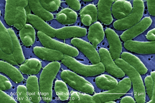
Vibrio vulnificus is a species of Gram-negative, motile, curved rod-shaped (vibrio), pathogenic bacteria of the genus Vibrio. Present in marine environments such as estuaries, brackish ponds, or coastal areas, V. vulnificus is related to V. cholerae, the causative agent of cholera. At least one strain of V. vulnificus is bioluminescent. Increasing seasonal ocean temperatures and low-salt marine environments like estuaries favor a greater concentration of Vibrio within filter-feeding shellfish; V. vulnificus infections in the Eastern United States have increased eightfold from 1988–2018.

<i>Haemophilus influenzae</i> Species of bacterium

Haemophilus influenzae is a Gram-negative, non-motile, coccobacillary, facultatively anaerobic, capnophilic pathogenic bacterium of the family Pasteurellaceae. The bacteria are mesophilic and grow best at temperatures between 35 and 37 °C.

An overwhelming post-splenectomy infection (OPSI) is a rare but rapidly fatal infection occurring in individuals following removal of the spleen. The infections are typically characterized by either meningitis or sepsis, and are caused by encapsulated organisms including Streptococcus pneumoniae. It is a medical emergency and requires immediate treatment. Death has been reported to occur within 12 hours.

<i>Neisseria meningitidis</i> Species of bacterium that can cause meningitis

Neisseria meningitidis, often referred to as the meningococcus, is a Gram-negative bacterium that can cause meningitis and other forms of meningococcal disease such as meningococcemia, a life-threatening sepsis. The bacterium is referred to as a coccus because it is round, and more specifically a diplococcus because of its tendency to form pairs.

<i>Moraxella catarrhalis</i> Species of bacterium

Moraxella catarrhalis is a fastidious, nonmotile, Gram-negative, aerobic, oxidase-positive diplococcus that can cause infections of the respiratory system, middle ear, eye, central nervous system, and joints of humans. It causes the infection of the host cell by sticking to the host cell using trimeric autotransporter adhesins.

<span class="mw-page-title-main">Meningococcal disease</span> Often life-threatening bacterial infection

Meningococcal disease describes infections caused by the bacterium Neisseria meningitidis. It has a high mortality rate if untreated but is vaccine-preventable. While best known as a cause of meningitis, it can also result in sepsis, which is an even more damaging and dangerous condition. Meningitis and meningococcemia are major causes of illness, death, and disability in both developed and under-developed countries.

<i>Eikenella corrodens</i> Species of bacterium

Eikenella corrodens is a Gram-negative facultative anaerobic bacillus that can cause severe invasive disease in humans. It was first identified by M. Eiken in 1958, who called it Bacteroides corrodens. E. corrodens is a rare pericarditis associated pathogen. It is a fastidious, slow growing, human commensal bacillus, capable of acting as an opportunistic pathogen and causing abscesses in several anatomical sites, including the liver, lung, spleen, and submandibular region. E. corrodens could independently cause serious infection in both immunocompetent and immunocompromised hosts.

<i>Elizabethkingia meningoseptica</i> Species of bacterium

Elizabethkingia meningoseptica is a Gram-negative, rod-shaped bacterium widely distributed in nature. It may be normally present in fish and frogs; it may be isolated from chronic infectious states, as in the sputum of cystic fibrosis patients. In 1959, American bacteriologist Elizabeth O. King was studying unclassified bacteria associated with pediatric meningitis at the Centers for Disease Control and Prevention in Atlanta, when she isolated an organism that she named Flavobacterium meningosepticum. In 1994, it was reclassified in the genus Chryseobacterium and renamed Chryseobacterium meningosepticum(chryseos = "golden" in Greek, so Chryseobacterium means a golden/yellow rod similar to Flavobacterium). In 2005, a 16S rRNA phylogenetic tree of Chryseobacteria showed that C. meningosepticum along with C. miricola were close to each other but outside the tree of the rest of the Chryseobacteria and were then placed in a new genus Elizabethkingia named after the original discoverer of F. meningosepticum.

<span class="mw-page-title-main">Ehrlichiosis</span> Medical condition

Ehrlichiosis is a tick-borne bacterial infection, caused by bacteria of the family Anaplasmataceae, genera Ehrlichia and Anaplasma. These obligate intracellular bacteria infect and kill white blood cells.

Pseudomonas oryzihabitans is a nonfermenting yellow-pigmented, gram-negative, rod-shaped bacterium that can cause sepsis, peritonitis, endophthalmitis, and bacteremia. It is an opportunistic pathogen of humans and warm-blooded animals that is commonly found in several environmental sources, from soil to rice paddies. They can be distinguished from other nonfermenters by their negative oxidase reaction and aerobic character. This organism can infect individuals that have major illnesses, including those undergoing surgery or with catheters in their body. Based on the 16S RNA analysis, these bacteria have been placed in the Pseudomonas putida group.

Capnocytophaga is a genus of Gram-negative bacteria. Normally found in the oropharyngeal tract of mammals, they are involved in the pathogenesis of some animal bite wounds and periodontal diseases.

<span class="mw-page-title-main">Pathogenic bacteria</span> Disease-causing bacteria

Pathogenic bacteria are bacteria that can cause disease. This article focuses on the bacteria that are pathogenic to humans. Most species of bacteria are harmless and are often beneficial but others can cause infectious diseases. The number of these pathogenic species in humans is estimated to be fewer than a hundred. By contrast, several thousand species are part of the gut flora present in the digestive tract.

Citrobacter koseri, formerly known as Citrobacter diversus, is a Gram-negative non-spore forming, rod-shaped bacterium. It is a facultative anaerobe capable of aerobic respiration. It is motile via peritrichous flagella. It is a member of the family of Enterobacteriaceae. The members of this family are part of the normal flora and commonly found in the digestive tracts of humans and animals. C. koseri may act as an opportunistic pathogen in individuals who are immunocompromised.

<span class="mw-page-title-main">Feline zoonosis</span> Medical condition

A feline zoonosis is a viral, bacterial, fungal, protozoan, nematode or arthropod infection that can be transmitted to humans from the domesticated cat, Felis catus. Some of these diseases are reemerging and newly emerging infections or infestations caused by zoonotic pathogens transmitted by cats. In some instances, the cat can display symptoms of infection and sometimes the cat remains asymptomatic. There can be serious illnesses and clinical manifestations in people who become infected. This is dependent on the immune status and age of the person. Those who live in close association with cats are more prone to these infections, but those that do not keep cats as pets can also acquire these infections as the transmission can be from cat feces and the parasites that leave their bodies.

References

  1. 1 2 3 4 Pers, C.; Gahrn-Hansen, B.; Frederiksen, W. (1996-07-01). "Capnocytophaga canimorsus Septicemia in Denmark, 1982-1995: Review of 39 Cases". Clinical Infectious Diseases. 23 (1): 71–75. doi:10.1093/clinids/23.1.71. ISSN   1058-4838. PMID   8816132.
  2. 1 2 3 4 5 Brenner, D J; Hollis, D G; Fanning, G R; Weaver, R E (1989). "Capnocytophaga canimorsus sp. nov. (formerly CDC group DF-2), a cause of septicemia following dog bite, and C. cynodegmi sp. nov., a cause of localized wound infection following dog bite". Journal of Clinical Microbiology. 27 (2): 231–235. doi:10.1128/jcm.27.2.231-235.1989. ISSN   0095-1137. PMC   267282 . PMID   2915017.
  3. 1 2 Fischer, L J; Weyant, R S; White, E H; Quinn, F D (1995). "Intracellular multiplication and toxic destruction of cultured macrophages by Capnocytophaga canimorsus". Infection and Immunity. 63 (9): 3484–3490. doi:10.1128/iai.63.9.3484-3490.1995. ISSN   0019-9567. PMC   173481 . PMID   7642281.
  4. 1 2 3 4 5 6 7 8 9 10 11 12 13 14 15 16 17 18 Lion, C.; Escande, F.; Burdin, J.C. (1996). "Capnocytophaga canimorsus infections in human: Review of the literature and cases report". European Journal of Epidemiology. 12 (5): 521–533. doi:10.1007/BF00144007. PMID   8905316. S2CID   22408402.
  5. Le Moal G; Landron C; Grollier G; Robert R; Burucoa C (2003). "Meningitis Due to Capnocytophaga canimorsus after Receipt of a Dog Bite: Case Report and Review of the Literature". Clin Infect Dis. 36 (3): e42–46. doi: 10.1086/345477 . PMID   12539089.
  6. Henry, Ronnie (2018). "Etymologia: Capnocytophaga canimorsus". Emerging Infectious Diseases. 24 (12): 2201. doi: 10.3201/eid2412.ET2412 . ISSN   1080-6040. PMC   6256413 .
  7. "Animal bites Fact sheet N°373". World Health Organization. February 2013. Archived from the original on 4 May 2015. Retrieved 5 May 2014.
  8. "Dog Bite Prevention". Centers for Disease Control and Prevention. Archived from the original on 1 May 2013. Retrieved 22 April 2013.
  9. 1 2 Janda, J. Michael; Graves, Margot H.; Lindquist, David; Probert, Will S. (2006). "Diagnosing Capnocytophaga canimorsus Infections". Emerging Infectious Diseases. Centers for Disease Control and Prevention (CDC). 12 (2): 340–342. doi:10.3201/eid1202.050783. ISSN   1080-6040. PMC   3373098 . PMID   16494769.
  10. 1 2 3 4 5 6 7 Gaastra, Wim; Lipman, Len J.A. (2010). "Capnocytophaga canimorsus". Veterinary Microbiology. Elsevier BV. 140 (3–4): 339–346. doi:10.1016/j.vetmic.2009.01.040. ISSN   0378-1135. PMID   19268498.
  11. 1 2 Renzi, Francesco; Ittig, Simon J.; Sadovskaya, Irina; Hess, Estelle; Lauber, Frederic; Dol, Melanie; Shin, Hwain; Mally, Manuela; Fiechter, Chantal; Sauder, Ursula; Chami, Mohamed; Cornelis, Guy R. (15 December 2016). "Evidence for a LOS and a capsular polysaccharide in Capnocytophaga canimorsus". Scientific Reports. 6: 38914. Bibcode:2016NatSR...638914R. doi: 10.1038/srep38914 . PMC   5156936 . PMID   27974829.
  12. 1 2 3 4 Shin, Hwain; Mally, Manuela; Meyer, Salome; Fiechter, Chantal; Paroz, Cécile; Zaehringer, Ulrich; Cornelis, Guy R. (2009). "Resistance of Capnocytophaga canimorsus to Killing by Human Complement and Polymorphonuclear Leukocytes". Infection and Immunity. 77 (6): 2262–2271. doi:10.1128/IAI.01324-08. ISSN   0019-9567. PMC   2687352 . PMID   19307219.
  13. de Boer, M.G.J.; Lambregts, P.C.L.A.; van Dam, A.P.; van ’t Wout, J.W. (2007). "Meningitis caused by Capnocytophaga canimorsus: When to expect the unexpected". Clinical Neurology and Neurosurgery. Elsevier BV. 109 (5): 393–398. doi:10.1016/j.clineuro.2007.02.010. ISSN   0303-8467. PMID   17408852. S2CID   8501012.
  14. Manfredi, P; Pagni, M; Cornelis, G. R. (2011). "Complete genome sequence of the dog commensal and human pathogen Capnocytophaga canimorsus strain 5". Journal of Bacteriology. 193 (19): 5558–9. doi:10.1128/JB.05853-11. PMC   3187460 . PMID   21914877.
  15. AW, Segal (2006). "How superoxide production by neutrophil leukocytes kills microbes". Novartis Foundation Symposium. Novartis Found Symp. 279: 92–8, discussion 98-100, 216–9. ISSN   1528-2511. PMID   17278388 . Retrieved 2024-01-23.
  16. Happel, K. I. (2005-12-01). "Alcohol, Immunosuppression, and the Lung". Proceedings of the American Thoracic Society. 2 (5): 428–432. doi:10.1513/pats.200507-065JS. ISSN   1546-3222. PMID   16322595.
  17. 1 2 Shin, Hwain; Mally, Manuela; Kuhn, Marina; Paroz, Cecile; Cornelis, Guy R. (2007). "Escape from Immune Surveillance by Capnocytophaga canimorsus". The Journal of Infectious Diseases. 195 (3): 375–386. doi:10.1086/510243. ISSN   0022-1899. PMID   17205476.