Related Research Articles

The Apicomplexa are a large phylum of parasitic alveolates. Most of them possess a unique form of organelle that comprises a type of non-photosynthetic plastid called an apicoplast, and an apical complex structure. The organelle is an adaptation that the apicomplexan applies in penetration of a host cell.

Plasmodium is a genus of unicellular eukaryotes that are obligate parasites of vertebrates and insects. The life cycles of Plasmodium species involve development in a blood-feeding insect host which then injects parasites into a vertebrate host during a blood meal. Parasites grow within a vertebrate body tissue before entering the bloodstream to infect red blood cells. The ensuing destruction of host red blood cells can result in malaria. During this infection, some parasites are picked up by a blood-feeding insect, continuing the life cycle.

The Plasmodiidae are a family of apicomplexan parasites, including the type genus Plasmodium, which is responsible for malaria. This family was erected in 1903 by Mesnil and is one of the four families in the order Haemospororida.

Eimeria tenella is a species of Eimeria that causes hemorrhagic cecal coccidiosis in young poultry. It is found worldwide.

Eimeria is a genus of apicomplexan parasites that includes various species capable of causing the disease coccidiosis in animals such as cattle, poultry and smaller ruminants including sheep and goats. Eimeria species are considered to be monoxenous because the life cycle is completed within a single host, and stenoxenous because they tend to be host specific, although a number of exceptions have been identified. Species of this genus infect a wide variety of hosts. Thirty-one species are known to occur in bats (Chiroptera), two in turtles, and 130 named species infect fish. Two species infect seals. Five species infect llamas and alpacas: E. alpacae, E. ivitaensis, E. lamae, E. macusaniensis, and E. punonensis. A number of species infect rodents, including E. couesii, E. kinsellai, E. palustris, E. ojastii and E. oryzomysi. Others infect poultry, rabbits and cattle. For full species list, see below.
The gregarines are a group of Apicomplexan alveolates, classified as the Gregarinasina or Gregarinia. The large parasites inhabit the intestines of many invertebrates. They are not found in any vertebrates. However, gregarines are closely related to both Toxoplasma and Plasmodium, which cause toxoplasmosis and malaria, respectively. Both protists use protein complexes similar to those that are formed by the gregarines for gliding motility and invading target cells. This makes them excellent models for studying gliding motility with the goal of developing treatment options for toxoplasmosis and malaria. Thousands of different species of gregarines are expected to be found in insects, and 99% of these gregarines still need to be described. Each insect can be the host of multiple species. One of the most studied gregarines is Gregarina garnhami. In general, gregarines are regarded as very successful parasites, as their hosts are spread over the entire planet.

Leucocytozoon is a genus of parasitic alveolates belonging to the phylum Apicomplexa.
Megaloschizonts are large schizonts that produce extremely high numbers of merozoites. They are found in various species of the Phylum Apicomplexa. The Apicomplexa phylum contains several parasitic protozoans. They have a very complex life cycle that includes several stages. Megaloschizonts and the smaller schizonts are the part of the life cycle that takes place inside the infected host organism and operates as an asexually reproductive cell. Megaloschizonts appear as grey-white nodules found in the smooth muscle of major organs, such as the heart, liver, lung or spleen.

Adeleorina is a suborder of parasites in the phylum Apicomplexa.
Karyolysus is a genus of coccidia. With the exception of K. sonomae whose vertebrate host is the yellow-legged frog, species in this genus only infect lizards of the genus Lacerta.
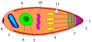
Apicomplexans, a group of intracellular parasites, have life cycle stages that allow them to survive the wide variety of environments they are exposed to during their complex life cycle. Each stage in the life cycle of an apicomplexan organism is typified by a cellular variety with a distinct morphology and biochemistry.
Acroeimeria is a genus of parasites that contains those species which initially develop immediately beneath the brush-border of the intestinal epithelium, but the meronts and gamonts of which are early on extruded to form a layer on the surface of the gut mucosa. Morphologically they are similar to the Eimeria to which they are closely related. The genus was described in 1989 by Paperna and Landsberg.
The genus Polychromophilus consists of obligate intracellular eukaryotic parasites that infect bats from every continent except Antarctica. They are transmitted by bat flies, which act as an insect vector as well as the parasite’s site of sporogeny. Polychromophilus follows a fairly typical Haemospororidian lifecycle, with gametocytes and gametes restricted to the bloodstream of the host and meronts infecting organs – most notably the lungs and the liver. The type species is Polychromophilus melanipherus, and was described by Dionisi in 1898.
Dactylosoma is a genus of parasitic alveolates of the phylum Apicomplexia.
Epieimeria is a genus of parasitic alveaolates of the phylum Apicomplexa.
Ovivora is a genus in the phylum Apicomplexa.
Merocystis is a genus in the phylum Apicomplexa.
Lankesterella is a genus in the phylum Apicomplexa. Species in this genus infect amphibians, reptiles and birds.
Mattesia is a genus of parasitic alveolates of the phylum Apicomplexa. Species in this genus infect insects.
Lipocystis is a genus of parasitic alveolates of the phylum Apicomplexa.
References
- ↑ Le Bail O, Landau I (1974) Description and experimental life cycle of Schellackia balli n. sp. (Lankesterellidae) a parasite of toads in Guyana. Ann Parasitol Hum Comp 49(6):663-668
- ↑ Finkelman, S.; Paperna, I. (2014). "Schellackia calotesi n. sp. from agamid lizards of the genus Calotes in Thailand". Parasite. 5 (1): 23–26. doi: 10.1051/parasite/1998051023 . ISSN 1252-607X. PMID 9754293.

- ↑ Lainson, R, Shaw JJ, Ward RD (1976) Schellackia landauae sp. nov. (Eimeriorina: Lankesterellidae) in the Brazilian lizard Polychrus marmoratus (Iguanidae): experimental transmission by Culex pipiens fatigans. Parasitol 11 (2)
- ↑ Bonorris, J.S., Ball, G.H.1955. Schellackia occidentalis n. sp., a blood-inhabiting coccidian found in lizards in Southern California. J Protozool 2: 31-34
- 1 2 3 BONORRIS, JIM S.; BALL, GORDON H. (February 1955). "Schellackia occidentalisn.sp., a Blood-inhabiting Coccidian Found in Lizards in Southern California". The Journal of Protozoology. 2 (1): 31–34. doi:10.1111/j.1550-7408.1955.tb02393.x. ISSN 0022-3921.
- 1 2 3 Megía-Palma, Rodrigo; Martínez, Javier; Cuervo, José J.; Jiménez-Robles, O.; Gomes, Verónica; Cabidof, C.; Fitzed, P.S.; Martina, J.; Merinoa, S. (2016). "Molecular diversity of the genus Schellackia (Apicomplexa: Schellackiidae) parasitizing lizards of the family lacertidae (squamata)". Molecular: 79.
- 1 2 3 4 5 6 7 Bristovetzky, Mariana; Paperna, Ilan (November 1990). "Life cycle and transmission of Schellackia cf. agamae, a parasite of the starred lizard Agama stellio". International Journal for Parasitology. 20 (7): 883–892. doi:10.1016/0020-7519(90)90026-j. ISSN 0020-7519.
- ↑ Paperna, I.; Lainson, R. (October 1995). "Schellackia (Apicomplexa: Eimeriidae) of the brazilian tree-frog, Phrynohyas venulosa (Amphibia: Anura) from Amazonian Brazil". Memórias do Instituto Oswaldo Cruz. 90 (5): 589–592. doi: 10.1590/s0074-02761995000500008 . ISSN 0074-0276.
- 1 2 3 Ostrovska, K.; Paperna, I. (1987). "Fine structure of gamont stages of Schellackia cf. agamae (Lankesterellidae, Eucoccidia) from the starred lizard Agama stellio". Parasitology Research. 73 (6): 492–499. doi:10.1007/bf00535322. ISSN 0044-3255.
- ↑ Paperna, I. (April 1993). "Electron microscopic study of Schellackia cf. agamae sporozoite infection in mosquitoes". International Journal for Parasitology. 23 (2): 187–190. doi:10.1016/0020-7519(93)90140-t. ISSN 0020-7519.
- ↑ Telford, Sam R. (June 1993). "A species of Schellackia (Apicomplexa: Lankesterellidae) parasitising east and southeast Asian lizards". Systematic Parasitology. 25 (2): 109–117. doi: 10.1007/bf00009980 . ISSN 0165-5752.