Visible spectrum

Last updated
White light is dispersed by a prism into the colors of the visible spectrum. Light dispersion of a mercury-vapor lamp with a flint glass prism IPNrdeg0125.jpg
White light is dispersed by a prism into the colors of the visible spectrum.

The visible spectrum is the band of the electromagnetic spectrum that is visible to the human eye. Electromagnetic radiation in this range of wavelengths is called visible light (or simply light). The optical spectrum is sometimes considered to be the same as the visible spectrum, but some authors define the term more broadly, to include the ultraviolet and infrared parts of the electromagnetic spectrum as well, known collectively as optical radiation . [1]

Contents

A typical human eye will respond to wavelengths from about 380 to about 750 nanometers. [2] In terms of frequency, this corresponds to a band in the vicinity of 400–790  terahertz. These boundaries are not sharply defined and may vary per individual. [3] Under optimal conditions these limits of human perception can extend to 310 nm (ultraviolet) and 1100 nm (near infrared). [4] [5] [6]

The spectrum does not contain all the colors that the human visual system can distinguish. Unsaturated colors such as pink, or purple variations like magenta, for example, are absent because they can only be made from a mix of multiple wavelengths. Colors containing only one wavelength are also called pure colors or spectral colors. [7]

Visible wavelengths pass largely unattenuated through the Earth's atmosphere via the "optical window" region of the electromagnetic spectrum. An example of this phenomenon is when clean air scatters blue light more than red light, and so the midday sky appears blue (apart from the area around the Sun which appears white because the light is not scattered as much). The optical window is also referred to as the "visible window" because it overlaps the human visible response spectrum. The near infrared (NIR) window lies just out of the human vision, as well as the medium wavelength infrared (MWIR) window, and the long-wavelength or far-infrared (LWIR or FIR) window, although other animals may perceive them.

Spectral colors

sRGB rendering of the spectrum of visible light Linear visible spectrum.svg
sRGB rendering of the spectrum of visible light
Color Wavelength
(nm)
Frequency
(THz)
Photon energy
(eV)
   violet
380–450670–7902.75–3.26
   blue
450–485620–6702.56–2.75
   cyan
485–500600–6202.48–2.56
   green
500–565530–6002.19–2.48
   yellow
565–590510–5302.10–2.19
   orange
590–625480–5101.98–2.10
   red
625–750400–4801.65–1.98

Colors that can be produced by visible light of a narrow band of wavelengths (monochromatic light) are called pure spectral colors. The various color ranges indicated in the illustration are an approximation: The spectrum is continuous, with no clear boundaries between one color and the next. [8]

History

Newton's color circle, from Opticks of 1704, showing the colors he associated with musical notes. The spectral colors from red to violet are divided by the notes of the musical scale, starting at D. The circle completes a full octave, from D to D. Newton's circle places red, at one end of the spectrum, next to violet, at the other. This reflects the fact that non-spectral purple colors are observed when red and violet light are mixed. Newton's color circle.png
Newton's color circle, from Opticks of 1704, showing the colors he associated with musical notes. The spectral colors from red to violet are divided by the notes of the musical scale, starting at D. The circle completes a full octave, from D to D. Newton's circle places red, at one end of the spectrum, next to violet, at the other. This reflects the fact that non-spectral purple colors are observed when red and violet light are mixed.

In the 13th century, Roger Bacon theorized that rainbows were produced by a similar process to the passage of light through glass or crystal. [9]

In the 17th century, Isaac Newton discovered that prisms could disassemble and reassemble white light, and described the phenomenon in his book Opticks . He was the first to use the word spectrum (Latin for "appearance" or "apparition") in this sense in print in 1671 in describing his experiments in optics. Newton observed that, when a narrow beam of sunlight strikes the face of a glass prism at an angle, some is reflected and some of the beam passes into and through the glass, emerging as different-colored bands. Newton hypothesized light to be made up of "corpuscles" (particles) of different colors, with the different colors of light moving at different speeds in transparent matter, red light moving more quickly than violet in glass. The result is that red light is bent (refracted) less sharply than violet as it passes through the prism, creating a spectrum of colors.

Newton's observation of prismatic colors (David Brewster 1855) Newton prismatic colours.JPG
Newton's observation of prismatic colors (David Brewster 1855)

Newton originally divided the spectrum into six named colors: red, orange, yellow, green, blue, and violet. He later added indigo as the seventh color since he believed that seven was a perfect number as derived from the ancient Greek sophists, of there being a connection between the colors, the musical notes, the known objects in the Solar System, and the days of the week. [10] The human eye is relatively insensitive to indigo's frequencies, and some people who have otherwise-good vision cannot distinguish indigo from blue and violet. For this reason, some later commentators, including Isaac Asimov, [11] have suggested that indigo should not be regarded as a color in its own right but merely as a shade of blue or violet. Evidence indicates that what Newton meant by "indigo" and "blue" does not correspond to the modern meanings of those color words. Comparing Newton's observation of prismatic colors with a color image of the visible light spectrum shows that "indigo" corresponds to what is today called blue, whereas his "blue" corresponds to cyan. [12] [13] [14]

In the 18th century, Johann Wolfgang von Goethe wrote about optical spectra in his Theory of Colours . Goethe used the word spectrum (Spektrum) to designate a ghostly optical afterimage, as did Schopenhauer in On Vision and Colors . Goethe argued that the continuous spectrum was a compound phenomenon. Where Newton narrowed the beam of light to isolate the phenomenon, Goethe observed that a wider aperture produces not a spectrum but rather reddish-yellow and blue-cyan edges with white between them. The spectrum appears only when these edges are close enough to overlap.

In the early 19th century, the concept of the visible spectrum became more definite, as light outside the visible range was discovered and characterized by William Herschel (infrared) and Johann Wilhelm Ritter (ultraviolet), Thomas Young, Thomas Johann Seebeck, and others. [15] Young was the first to measure the wavelengths of different colors of light, in 1802. [16]

The connection between the visible spectrum and color vision was explored by Thomas Young and Hermann von Helmholtz in the early 19th century. Their theory of color vision correctly proposed that the eye uses three distinct receptors to perceive color.

Limits to visible range

Photopic (black) and scotopic (green) luminous efficiency functions. The horizontal axis is wavelength in nm. See luminous efficiency function for more info. Luminosity.svg
Photopic (black) and scotopic (green) luminous efficiency functions. The horizontal axis is wavelength in nm. See luminous efficiency function for more info.

The visible spectrum is limited to wavelengths that can both reach the retina and trigger visual phototransduction (excite a visual opsin). Insensitivity to UV light is generally limited by transmission through the lens. Insensitivity to IR light is limited by the spectral sensitivity functions of the visual opsins. The range is defined psychometrically by the luminous efficiency function, which accounts for all of these factors. In humans, there is a separate function for each of two visual systems, one for photopic vision, used in daylight, which is mediated by cone cells, and one for scotopic vision, used in dim light, which is mediated by rod cells. Each of these functions have different visible ranges. However, discussion on the visible range generally assumes photopic vision.

Atmospheric transmission

The visible range of most animals evolved to match the optical window, which is the range of light that can pass through the atmosphere. The ozone layer absorbs almost all UV light (below 315 nm). [17] However, this only affects cosmic light (e.g. sunlight), not terrestrial light (e.g. Bioluminescence).

Ocular transmission

Cumulative transmission spectra of light as it passes through the ocular media, namely after the cornea (blue), before the lens (red), after the lens (gray) and before the retina (orange). The solid lines are for a 4.5 year old eye. The dashed orange line is for a 53 year old eye, and dotted for a 75 year old eye, indicating the effect of lens yellowing.) Lens Cornea Transmission.png
Cumulative transmission spectra of light as it passes through the ocular media, namely after the cornea (blue), before the lens (red), after the lens (gray) and before the retina (orange). The solid lines are for a 4.5 year old eye. The dashed orange line is for a 53 year old eye, and dotted for a 75 year old eye, indicating the effect of lens yellowing.)

Before reaching the retina, light must first transmit through the cornea and lens. UVB light (< 315 nm) is filtered mostly by the cornea, and UVA light (315–400 nm) is filtered mostly by the lens. [18] The lens also yellows with age, attenuating transmission most strongly at the blue part of the spectrum. [18] This can cause xanthopsia as well as a slight truncation of the short-wave (blue) limit of the visible spectrum. Subjects with aphakia are missing a lens, so UVA light can reach the retina and excite the visual opsins; this expands the visible range and may also lead to cyanopsia.

Opsin absorption

Each opsin has a spectral sensitivity function, which defines how likely it is to absorb a photon of each wavelength. The luminous efficiency function is approximately the superposition of the contributing visual opsins. Variance in the position of the individual opsin spectral sensitivity functions therefore affects the luminous efficiency function and the visible range. For example, the long-wave (red) limit changes proportionally to the position of the L-opsin. The positions are defined by the peak wavelength (wavelength of highest sensitivity), so as the L-opsin peak wavelength blue shifts by 10 nm, the long-wave limit of the visible spectrum also shifts 10 nm. Large deviations of the L-opsin peak wavelength lead to a form of color blindness called protanomaly and a missing L-opsin (protanopia) shortens the visible spectrum by about 30 nm at the long-wave limit. Forms of color blindness affecting the M-opsin and S-opsin do not significantly affect the luminous efficiency function nor the limits of the visible spectrum.

Different definitions

Regardless of actual physical and biological variance, the definition of the limits is not standard and will change depending on the industry. For example, some industries may be concerned with practical limits, so would conservatively report 420–680 nm, [19] [20] while others may be concerned with psychometrics and achieving the broadest spectrum would liberally report 380–750, or even 380–800 nm. [21] [22] The luminous efficiency function in the NIR does not have a hard cutoff, but rather an exponential decay, such that the function's value (or vision sensitivity) at 1,050 nm is about 109 times weaker than at 700 nm; much higher intensity is therefore required to perceive 1,050 nm light than 700 nm light. [23]

Vision outside the visible spectrum

Under ideal laboratory conditions, subjects may perceive infrared light up to at least 1,064 nm. [23] While 1,050 nm NIR light can evoke red, suggesting direct absorption by the L-opsin, there are also reports that pulsed NIR lasers can evoke green, which suggests two-photon absorption may be enabling extended NIR sensitivity. [23]

Similarly, young subjects may perceive ultraviolet wavelengths down to about 310–313 nm, [24] [25] [26] but detection of light below 380 nm may be due to fluorescence of the ocular media, rather than direct absorption of UV light by the opsins. As UVA light is absorbed by the ocular media (lens and cornea), it may fluoresce and be released at a lower energy (longer wavelength) that can then be absorbed by the opsins. For example, when the lens absorbs 350 nm light, the fluorescence emission spectrum is centered on 440 nm. [27]

Non-visual light detection

In addition to the photopic and scotopic systems, humans have other systems for detecting light that do not contribute to the primary visual system. For example, melanopsin has an absorption range of 420–540 nm and regulates circadian rhythm and other reflexive processes. [28] Since the melanopsin system does not form images, it is not strictly considered vision and does not contribute to the visible range.

In non-humans

The visible spectrum is defined as that visible to humans, but the variance between species is large. Not only can cone opsins be spectrally shifted to alter the visible range, but vertebrates with 4 cones (tetrachromatic) or 2 cones (dichromatic) relative to humans' 3 (trichromatic) will also tend to have a wider or narrower visible spectrum than humans, respectively.

Vertebrates tend to have 1-4 different opsin classes: [17]

Testing the visual systems of animals behaviorally is difficult, so the visible range of animals is usually estimated by comparing the peak wavelengths of opsins with those of typical humans (S-opsin at 420 nm and L-opsin at 560 nm).

Mammals

Most mammals have retained only two opsin classes (LWS and VS), due likely to the nocturnal bottleneck. However, old world primates (including humans) have since evolved two versions in the LWS class to regain trichromacy. [17] Unlike most mammals, rodents' UVS opsins have remained at shorter wavelengths. Along with their lack of UV filters in the lens, mice have a UVS opsin that can detect down to 340 nm. While allowing UV light to reach the retina can lead to retinal damage, the short lifespan of mice compared with other mammals may minimize this disadvantage relative to the advantage of UV vision. [29] Dogs have two cone opsins at 429 nm and 555 nm, so see almost the entire visible spectrum of humans, despite being dichromatic. [30] Horses have two cone opsins at 428 nm and 539 nm, yielding a slightly more truncated red vision. [31]

Birds

Most other vertebrates (birds, lizards, fish, etc.) have retained their tetrachromacy, including UVS opsins that extend further into the ultraviolet than humans' VS opsin. [17] The sensitivity of avian UVS opsins vary greatly, from 355–425 nm, and LWS opsins from 560–570 nm. [32] This translates to some birds with a visible spectrum on par with humans, and other birds with greatly expanded sensitivity to UV light. The LWS opsin of birds is sometimes reported to have a peak wavelength above 600 nm, but this is an effective peak wavelength that incorporates the filter of avian oil droplets. [32] The peak wavelength of the LWS opsin alone is the better predictor of the long-wave limit. A possible benefit of avian UV vision involves sex-dependent markings on their plumage that are visible only in the ultraviolet range. [33] [34]

Fish

Teleosts (bony fish) are generally tetrachromatic. The sensitivity of fish UVS opsins vary from 347-383 nm, and LWS opsins from 500-570 nm. [35] However, some fish that use alternative chromophores can extend their LWS opsin sensitivity to 625 nm. [35] The popular belief that the common goldfish is the only animal that can see both infrared and ultraviolet light [36] is incorrect, because goldfish cannot see infrared light. [37]

Invertebrates

The visual systems of invertebrates deviate greatly from vertebrates, so direct comparisons are difficult. However, UV sensitivity has been reported in most insect species. [38] Bees and many other insects can detect ultraviolet light, which helps them find nectar in flowers. Plant species that depend on insect pollination may owe reproductive success to their appearance in ultraviolet light rather than how colorful they appear to humans. Bees' long-wave limit is at about 590 nm. [39] Mantis shrimp exhibit up to 14 opsins, enabling a visible range of less than 300 nm to above 700 nm. [17]

Thermal vision

Some snakes can "see" [40] radiant heat at wavelengths between 5 and 30  μm to a degree of accuracy such that a blind rattlesnake can target vulnerable body parts of the prey at which it strikes, [41] and other snakes with the organ may detect warm bodies from a meter away. [42] It may also be used in thermoregulation and predator detection. [43] [44]

Spectroscopy

Earth's atmosphere partially or totally blocks some wavelengths of electromagnetic radiation, but in visible light it is mostly transparent Atmospheric electromagnetic opacity.svg
Earth's atmosphere partially or totally blocks some wavelengths of electromagnetic radiation, but in visible light it is mostly transparent

Spectroscopy is the study of objects based on the spectrum of color they emit, absorb or reflect. Visible-light spectroscopy is an important tool in astronomy (as is spectroscopy at other wavelengths), where scientists use it to analyze the properties of distant objects. Chemical elements and small molecules can be detected in astronomical objects by observing emission lines and absorption lines. For example, Helium was first detected by analysis of the spectrum of the Sun. The shift in frequency of spectral lines is used to measure the Doppler shift (redshift or blueshift) of distant objects to determine their velocities towards or away from the observer. Astronomical spectroscopy uses high-dispersion diffraction gratings to observe spectra at very high spectral resolutions.

See also

Related Research Articles

<span class="mw-page-title-main">Color</span> Visual perception of the light spectrum

Color or colour is the visual perception based on the electromagnetic spectrum. Though color is not an inherent property of matter, color perception is related to an object's light absorption, reflection, emission spectra and interference. For most humans, colors are perceived in the visible light spectrum with three types of cone cells (trichromacy). Other animals may have a different number of cone cell types or have eyes sensitive to different wavelength, such as bees that can distinguish ultraviolet, and thus have a different color sensitivity range. Animal perception of color originates from different light wavelength or spectral sensitivity in cone cell types, which is then processed by the brain.

<span class="mw-page-title-main">Electromagnetic spectrum</span> Range of frequencies or wavelengths of electromagnetic radiation

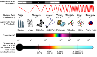
The electromagnetic spectrum is the full range of electromagnetic radiation, organized by frequency or wavelength. The spectrum is divided into separate bands, with different names for the electromagnetic waves within each band. From low to high frequency these are: radio waves, microwaves, infrared, visible light, ultraviolet, X-rays, and gamma rays. The electromagnetic waves in each of these bands have different characteristics, such as how they are produced, how they interact with matter, and their practical applications.

<span class="mw-page-title-main">Ultraviolet</span> Energetic, invisible light energy range

Ultraviolet (UV) light is electromagnetic radiation of wavelengths of 10–400 nanometers, shorter than that of visible light, but longer than X-rays. UV radiation is present in sunlight, and constitutes about 10% of the total electromagnetic radiation output from the Sun. It is also produced by electric arcs, Cherenkov radiation, and specialized lights, such as mercury-vapor lamps, tanning lamps, and black lights.

<span class="mw-page-title-main">Night vision</span> Ability to see in low light conditions

Night vision is the ability to see in low-light conditions, either naturally with scotopic vision or through a night-vision device. Night vision requires both sufficient spectral range and sufficient intensity range. Humans have poor night vision compared to many animals such as cats, dogs, foxes and rabbits, in part because the human eye lacks a tapetum lucidum, tissue behind the retina that reflects light back through the retina thus increasing the light available to the photoreceptors.

<span class="mw-page-title-main">Color vision</span> Ability to perceive differences in light frequency

Color vision, a feature of visual perception, is an ability to perceive differences between light composed of different frequencies independently of light intensity.

<span class="mw-page-title-main">Photometry (optics)</span> Science of the measurement of visible light

Photometry is a branch of science that deals with the measurement of light in terms of its perceived brightness to the human eye. It is concerned with quantifying the amount of light that is emitted, transmitted, or received by an object or a system.

<span class="mw-page-title-main">Spectrophotometry</span> Branch of spectroscopy

Spectrophotometry is a branch of electromagnetic spectroscopy concerned with the quantitative measurement of the reflection or transmission properties of a material as a function of wavelength. Spectrophotometry uses photometers, known as spectrophotometers, that can measure the intensity of a light beam at different wavelengths. Although spectrophotometry is most commonly applied to ultraviolet, visible, and infrared radiation, modern spectrophotometers can interrogate wide swaths of the electromagnetic spectrum, including x-ray, ultraviolet, visible, infrared, and/or microwave wavelengths.

<span class="mw-page-title-main">Tetrachromacy</span> Type of color vision with four types of cone cells

Tetrachromacy is the condition of possessing four independent channels for conveying color information, or possessing four types of cone cell in the eye. Organisms with tetrachromacy are called tetrachromats.

<span class="mw-page-title-main">Trichromacy</span> Possessing of three independent channels for conveying color information

Trichromacy or trichromatism is the possession of three independent channels for conveying color information, derived from the three different types of cone cells in the eye. Organisms with trichromacy are called trichromats.

<span class="mw-page-title-main">Cone cell</span> Photoreceptor cells responsible for color vision made to function in bright light

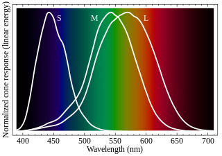
Cone cells or cones are photoreceptor cells in the retinas of vertebrates' eyes. They respond differently to light of different wavelengths, and the combination of their responses is responsible for color vision. Cones function best in relatively bright light, called the photopic region, as opposed to rod cells, which work better in dim light, or the scotopic region. Cone cells are densely packed in the fovea centralis, a 0.3 mm diameter rod-free area with very thin, densely packed cones which quickly reduce in number towards the periphery of the retina. Conversely, they are absent from the optic disc, contributing to the blind spot. There are about six to seven million cones in a human eye, with the highest concentration being towards the macula.

<span class="mw-page-title-main">Spectral color</span> Color evoked by a single wavelength of light in the visible spectrum

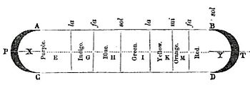
A spectral color is a color that is evoked by monochromatic light, i.e. either a spectral line with a single wavelength or frequency of light in the visible spectrum, or a relatively narrow spectral band. Every wave of visible light is perceived as a spectral color; when viewed as a continuous spectrum, these colors are seen as the familiar rainbow. Non-spectral colors are evoked by a combination of spectral colors.

A spectroradiometer is a light measurement tool that is able to measure both the wavelength and amplitude of the light emitted from a light source. Spectrometers discriminate the wavelength based on the position the light hits at the detector array allowing the full spectrum to be obtained with a single acquisition. Most spectrometers have a base measurement of counts which is the un-calibrated reading and is thus impacted by the sensitivity of the detector to each wavelength. By applying a calibration, the spectrometer is then able to provide measurements of spectral irradiance, spectral radiance and/or spectral flux. This data is also then used with built in or PC software and numerous algorithms to provide readings or Irradiance (W/cm2), Illuminance, Radiance (W/sr), Luminance (cd), Flux, Chromaticity, Color Temperature, Peak and Dominant Wavelength. Some more complex spectrometer software packages also allow calculation of PAR μmol/m2/s, Metamerism, and candela calculations based on distance and include features like 2- and 20-degree observer, baseline overlay comparisons, transmission and reflectance.

<span class="mw-page-title-main">Opsin</span> Class of light-sensitive proteins

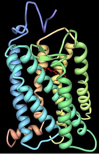
Animal opsins are G-protein-coupled receptors and a group of proteins made light-sensitive via a chromophore, typically retinal. When bound to retinal, opsins become retinylidene proteins, but are usually still called opsins regardless. Most prominently, they are found in photoreceptor cells of the retina. Five classical groups of opsins are involved in vision, mediating the conversion of a photon of light into an electrochemical signal, the first step in the visual transduction cascade. Another opsin found in the mammalian retina, melanopsin, is involved in circadian rhythms and pupillary reflex but not in vision. Humans have in total nine opsins. Beside vision and light perception, opsins may also sense temperature, sound, or chemicals.

<span class="mw-page-title-main">Ultraviolet photography</span> Photographic process using UV radiation

Ultraviolet photography is a photographic process of recording images by using radiation from the ultraviolet (UV) spectrum only. Images taken with ultraviolet radiation serve a number of scientific, medical or artistic purposes. Images may reveal deterioration of art works or structures not apparent under light. Diagnostic medical images may be used to detect certain skin disorders or as evidence of injury. Some animals, particularly insects, use ultraviolet wavelengths for vision; ultraviolet photography can help investigate the markings of plants that attract insects, while invisible to the unaided human eye. Ultraviolet photography of archaeological sites may reveal artifacts or traffic patterns not otherwise visible.

<span class="mw-page-title-main">OPN1LW</span> Protein-coding gene in humans

OPN1LW is a gene on the X chromosome that encodes for long wave sensitive (LWS) opsin, or red cone photopigment. It is responsible for perception of visible light in the yellow-green range on the visible spectrum. The gene contains 6 exons with variability that induces shifts in the spectral range. OPN1LW is subject to homologous recombination with OPN1MW, as the two have very similar sequences. These recombinations can lead to various vision problems, such as red-green colourblindness and blue monochromacy. The protein encoded is a G-protein coupled receptor with embedded 11-cis-retinal, whose light excitation causes a cis-trans conformational change that begins the process of chemical signalling to the brain.

<span class="mw-page-title-main">Evolution of color vision in primates</span> Loss and regain of colour vision during the evolution of primates

The evolution of color vision in primates is highly unusual compared to most eutherian mammals. A remote vertebrate ancestor of primates possessed tetrachromacy, but nocturnal, warm-blooded, mammalian ancestors lost two of four cones in the retina at the time of dinosaurs. Most teleost fish, reptiles and birds are therefore tetrachromatic while most mammals are strictly dichromats, the exceptions being some primates and marsupials, who are trichromats, and many marine mammals, who are monochromats.

<span class="mw-page-title-main">Bird vision</span> Senses for birds

Vision is the most important sense for birds, since good eyesight is essential for safe flight. Birds have a number of adaptations which give visual acuity superior to that of other vertebrate groups; a pigeon has been described as "two eyes with wings". Birds are theropod dinosaurs, and the avian eye resembles that of other reptiles, with ciliary muscles that can change the shape of the lens rapidly and to a greater extent than in the mammals. Birds have the largest eyes relative to their size in the animal kingdom, and movement is consequently limited within the eye's bony socket. In addition to the two eyelids usually found in vertebrates, bird's eyes are protected by a third transparent movable membrane. The eye's internal anatomy is similar to that of other vertebrates, but has a structure, the pecten oculi, unique to birds.

<span class="mw-page-title-main">Full-spectrum photography</span> Photography capturing visible and near-infrared light

Full-spectrum photography is a subset of multispectral imaging, defined among photography enthusiasts as imaging with consumer cameras the full, broad spectrum of a film or camera sensor bandwidth. In practice, specialized broadband/full-spectrum film captures visible and near infrared light, commonly referred to as the "VNIR".

<span class="mw-page-title-main">Congenital red–green color blindness</span> Most common genetic condition leading to color blindness

Congenital red–green color blindness is an inherited condition that is the root cause of the majority of cases of color blindness. It has no significant symptoms aside from its minor to moderate effect on color vision. It is caused by variation in the functionality of the red and/or green opsin proteins, which are the photosensitive pigment in the cone cells of the retina, which mediate color vision. Males are more likely to inherit red–green color blindness than females, because the genes for the relevant opsins are on the X chromosome. Screening for congenital red–green color blindness is typically performed with the Ishihara or similar color vision test. There is no cure for color blindness.

<span class="mw-page-title-main">Vertebrate visual opsin</span>

Vertebrate visual opsins are a subclass of ciliary opsins and mediate vision in vertebrates. They include the opsins in human rod and cone cells. They are often abbreviated to opsin, as they were the first opsins discovered and are still the most widely studied opsins.

References

  1. Pedrotti, Frank L.; Pedrotti, Leno M.; Pedrotti, Leno S. (December 21, 2017). Introduction to Optics. Cambridge University Press. pp. 7–8. ISBN   9781108428262.
  2. Starr, Cecie (2005). Biology: Concepts and Applications . Thomson Brooks/Cole. p.  94. ISBN   978-0-534-46226-0.
  3. "The visible spectrum". Britannica.
  4. D. H. Sliney (February 2016). "What is light? The visible spectrum and beyond". Eye. 30 (2): 222–229. doi:10.1038/eye.2015.252. ISSN   1476-5454. PMC   4763133 . PMID   26768917.
  5. W. C. Livingston (2001). Color and light in nature (2nd ed.). Cambridge, UK: Cambridge University Press. ISBN   0-521-77284-2.
  6. Grazyna Palczewska; et al. (December 2014). "Human infrared vision is triggered by two-photon chromophore isomerization". Proceedings of the National Academy of Sciences. 111 (50): E5445–E5454. Bibcode:2014PNAS..111E5445P. doi: 10.1073/pnas.1410162111 . PMC   4273384 . PMID   25453064.
  7. Nave, R. "Spectral Colors". Hyperphysics. Retrieved 2022-05-11.
  8. Bruno, Thomas J. and Svoronos, Paris D. N. (2005). CRC Handbook of Fundamental Spectroscopic Correlation Charts. CRC Press. ISBN   9781420037685
  9. Coffey, Peter (1912). The Science of Logic: An Inquiry Into the Principles of Accurate Thought. Longmans. p.  185. roger bacon prism.
  10. Isacoff, Stuart (16 January 2009). Temperament: How Music Became a Battleground for the Great Minds of Western Civilization. Knopf Doubleday Publishing Group. pp. 12–13. ISBN   978-0-307-56051-3 . Retrieved 18 March 2014.
  11. Asimov, Isaac (1975). Eyes on the universe : a history of the telescope . Boston: Houghton Mifflin. p.  59. ISBN   978-0-395-20716-1.
  12. Evans, Ralph M. (1974). The perception of color (null ed.). New York: Wiley-Interscience. ISBN   978-0-471-24785-2.
  13. McLaren, K. (March 2007). "Newton's indigo". Color Research & Application. 10 (4): 225–229. doi:10.1002/col.5080100411.
  14. Waldman, Gary (2002). Introduction to light : the physics of light, vision, and color (Dover ed.). Mineola: Dover Publications. p. 193. ISBN   978-0-486-42118-6.
  15. Mary Jo Nye, ed. (2003). The Cambridge History of Science: The Modern Physical and Mathematical Sciences. Vol. 5. Cambridge University Press. p. 278. ISBN   978-0-521-57199-9.
  16. John C. D. Brand (1995). Lines of light: the sources of dispersive spectroscopy, 1800–1930. CRC Press. pp. 30–32. ISBN   978-2-88449-163-1.
  17. 1 2 3 4 5 Hunt, D.M.; Wilkie, S.E.; Bowmaker, J.K.; Poopalasundaram, S. (October 2001). "Vision in the ultraviolet". Cellular and Molecular Life Sciences. 58 (11): 1583–1598. doi:10.1007/PL00000798. PMID   11706986. S2CID   22938704. Radiation below 320 nm [ultraviolet (UV)A] is largely screened out by the ozone layer in the Earth's upper atmosphere and is therefore unavailable to the visual system,
  18. 1 2 Boettner, Edward A.; Wolter, J. Reimer (December 1962). "Transmission of Ocular Media". Investigative Ophthalmology & Visual Science. 1: 776-783.
  19. Laufer, Gabriel (1996). "Geometrical Optics". Introduction to Optics and Lasers in Engineering. p. 11. Bibcode:1996iole.book.....L. doi:10.1017/CBO9781139174190.004. ISBN   978-0-521-45233-5 . Retrieved 20 October 2013.
  20. Bradt, Hale (2004). Astronomy Methods: A Physical Approach to Astronomical Observations. Cambridge University Press. p. 26. ISBN   978-0-521-53551-9 . Retrieved 20 October 2013.
  21. Ohannesian, Lena; Streeter, Anthony (2001). Handbook of Pharmaceutical Analysis. CRC Press. p. 187. ISBN   978-0-8247-4194-5 . Retrieved 20 October 2013.
  22. Ahluwalia, V.K.; Goyal, Madhuri (2000). A Textbook of Organic Chemistry. Narosa. p. 110. ISBN   978-81-7319-159-6 . Retrieved 20 October 2013.
  23. 1 2 3 Sliney, David H.; Wangemann, Robert T.; Franks, James K.; Wolbarsht, Myron L. (1976). "Visual sensitivity of the eye to infrared laser radiation". Journal of the Optical Society of America . 66 (4): 339–341. Bibcode:1976JOSA...66..339S. doi:10.1364/JOSA.66.000339. PMID   1262982. The foveal sensitivity to several near-infrared laser wavelengths was measured. It was found that the eye could respond to radiation at wavelengths at least as far as 1,064 nm. A continuous 1,064 nm laser source appeared red, but a 1,060 nm pulsed laser source appeared green, which suggests the presence of second harmonic generation in the retina.
  24. Lynch, David K.; Livingston, William Charles (2001). Color and Light in Nature (2nd ed.). Cambridge: Cambridge University Press. p. 231. ISBN   978-0-521-77504-5. Archived from the original on 8 October 2022. Retrieved 12 October 2013. Limits of the eye's overall range of sensitivity extends from about 310 to 1,050 nanometers
  25. Dash, Madhab Chandra; Dash, Satya Prakash (2009). Fundamentals of Ecology 3E. Tata McGraw-Hill Education. p. 213. ISBN   978-1-259-08109-5. Archived from the original on 8 October 2022. Retrieved 18 October 2013. Normally the human eye responds to light rays from 390 to 760 nm. This can be extended to a range of 310 to 1,050 nm under artificial conditions.
  26. Saidman, Jean (15 May 1933). "Sur la visibilité de l'ultraviolet jusqu'à la longueur d'onde 3130" [The visibility of the ultraviolet to the wave length of 3130]. Comptes rendus de l'Académie des sciences (in French). 196: 1537–9. Archived from the original on 24 October 2013. Retrieved 21 October 2013.
  27. Kurzel, Richard B.; Wolbarsht, Myron L.; Yamanashi, Bill S. (1977). "Ultraviolet Radiation Effects on the Human Eye". Photochemical and Photobiological Reviews. pp. 133–167. doi:10.1007/978-1-4684-2577-2_3. ISBN   978-1-4684-2579-6.
  28. Enezi J, Revell V, Brown T, Wynne J, Schlangen L, Lucas R (August 2011). "A "melanopic" spectral efficiency function predicts the sensitivity of melanopsin photoreceptors to polychromatic lights". Journal of Biological Rhythms. 26 (4): 314–323. doi: 10.1177/0748730411409719 . PMID   21775290. S2CID   22369861.
  29. Gouras, Peter; Ekesten, Bjorn (December 2004). "Why do mice have ultra-violet vision?". Experimental Eye Research. 79 (6): 887–892. doi:10.1016/j.exer.2004.06.031. PMID   15642326.
  30. Neitz, Jay; Geist, Timothy; Jacobs, Gerald H. (August 1989). "Color vision in the dog". Visual Neuroscience. 3 (2): 119–125. doi:10.1017/S0952523800004430. PMID   2487095. S2CID   23509491.
  31. Carroll, Joseph; Murphy, Christopher J.; Neitz, Maureen; Ver Hoeve, James N.; Neitz, Jay (3 October 2001). "Photopigment basis for dichromatic color vision in the horse". Journal of Vision. 1 (2): 80–87. doi: 10.1167/1.2.2 . PMID   12678603.
  32. 1 2 Hart, Nathan S.; Hunt, David M. (January 2007). "Avian Visual Pigments: Characteristics, Spectral Tuning, and Evolution". The American Naturalist. 169 (S1): S7–S26. doi:10.1086/510141. PMID   19426092. S2CID   25779190.
  33. Cuthill, Innes C (1997). "Ultraviolet vision in birds". In Peter J.B. Slater (ed.). Advances in the Study of Behavior. Vol. 29. Oxford, England: Academic Press. p. 161. ISBN   978-0-12-004529-7.
  34. Jamieson, Barrie G. M. (2007). Reproductive Biology and Phylogeny of Birds. Charlottesville VA: University of Virginia. p. 128. ISBN   978-1-57808-386-2.
  35. 1 2 Carleton, Karen L.; Escobar-Camacho, Daniel; Stieb, Sara M.; Cortesi, Fabio; Marshall, N. Justin (15 April 2020). "Seeing the rainbow: mechanisms underlying spectral sensitivity in teleost fishes". Journal of Experimental Biology. 223 (8). doi:10.1242/jeb.193334. PMC   7188444 . PMID   32327561.
  36. "True or False? "The common goldfish is the only animal that can see both infra-red and ultra-violet light."". Skeptive. 2013. Archived from the original on December 24, 2013. Retrieved September 28, 2013.
  37. Neumeyer, Christa (2012). "Chapter 2: Color Vision in Goldfish and Other Vertebrates". In Lazareva, Olga; Shimizu, Toru; Wasserman, Edward (eds.). How Animals See the World: Comparative Behavior, Biology, and Evolution of Vision. Oxford Scholarship Online. ISBN   978-0-19-533465-4.
  38. Briscoe, Adriana D.; Chittka, Lars (January 2001). "T<SCP>HE</SCP>E<SCP>VOLUTION OF</SCP>C<SCP>OLOR</SCP>V<SCP>ISION IN</SCP>I<SCP>NSECTS</SCP>". Annual Review of Entomology. 46 (1): 471–510. doi:10.1146/annurev.ento.46.1.471. PMID   11112177.
  39. Skorupski, Peter; Chittka, Lars (10 August 2010). "Photoreceptor Spectral Sensitivity in the Bumblebee, Bombus impatiens (Hymenoptera: Apidae)". PLOS ONE. 5 (8): e12049. Bibcode:2010PLoSO...512049S. doi: 10.1371/journal.pone.0012049 . PMC   2919406 . PMID   20711523.
  40. Newman, EA; Hartline, PH (1981). "Integration of visual and infrared information in bimodal neurons in the rattlesnake optic tectum". Science. 213 (4509): 789–91. Bibcode:1981Sci...213..789N. doi:10.1126/science.7256281. PMC   2693128 . PMID   7256281.
  41. Kardong, KV; Mackessy, SP (1991). "The strike behavior of a congenitally blind rattlesnake". Journal of Herpetology. 25 (2): 208–211. doi:10.2307/1564650. JSTOR   1564650.
  42. Fang, Janet (14 March 2010). "Snake infrared detection unravelled". Nature News. doi:10.1038/news.2010.122.
  43. Krochmal, Aaron R.; George S. Bakken; Travis J. LaDuc (15 November 2004). "Heat in evolution's kitchen: evolutionary perspectives on the functions and origin of the facial pit of pitvipers (Viperidae: Crotalinae)". Journal of Experimental Biology. 207 (Pt 24): 4231–4238. doi: 10.1242/jeb.01278 . PMID   15531644.
  44. Greene HW. (1992). "The ecological and behavioral context for pitviper evolution", in Campbell JA, Brodie ED Jr. Biology of the Pitvipers. Texas: Selva. ISBN   0-9630537-0-1.