Darier's disease

Last updated
Darier's disease
Other namesDarier disease, Darier–White disease, [1] Dyskeratosis follicularis, [1] and Keratosis follicularis [2] :523 [3] :567
Darier's disease.jpg
Linear Darier's disease
Specialty Medical genetics   OOjs UI icon edit-ltr-progressive.svg

Darier's disease (DAR) is a rare, inherited skin disorder that presents with multiple greasy, crusting, thick brown bumps that merge into patches. [4] It is an autosomal dominant disorder discovered by French dermatologist Ferdinand-Jean Darier.

Contents

Mild forms of the disease are the most common, consisting solely of skin rashes that flare up under certain conditions such as high humidity, high stress, or tight-fitting clothes. Short stature, when combined with poorly-formed fingernails that contain vertical striations, is diagnostic even for mild forms of DAR. Symptoms will usually appear in late childhood or early adulthood between the ages of 15 and 30 years and will vary over the lifespan.

More severe cases are characterized by dark crusty patches on the skin that are mildly greasy and that emit a strong odor. These patches, also known as keratotic papules, keratosis follicularis, or dyskeratosis follicularis, most often appear on the scalp, forehead, upper arms, chest, back, knees, elbows, and behind the ear. [5] [6]

Signs and symptoms

Genetics

Mutations in a single gene, ATP2A2, are responsible for the development of Darier's disease. ATP2A2 encodes the SERCA2 protein, which is a calcium pump localized to the membranes of the endoplasmic reticulum (ER) in nearly all cells and the sarcoplasmic reticulum (SR) in muscle cells. The ER is where protein processing and transport begins for proteins targeted for secretion. The SR is a specialized form of ER found in muscle cells that sequesters calcium, the regulated efflux of which into the cytosol stimulates muscle fiber contraction. Calcium acts as a second messenger in many cellular signal transduction pathways. SERCA2 is required for Ca2+ signaling in cells by removing nearly all Ca2+ ions from the cytoplasm and storing them in the ER/SR compartments. [10] [11] [12] [13]

Darier's disease has an autosomal dominant pattern of inheritance. Autosomal dominant - en.svg
Darier's disease has an autosomal dominant pattern of inheritance.

A large number of mutant alleles of ATP2A2 have been identified in association with Darier's Disease. One study of 19 families and 6 sporadic cases found 24 specific, novel mutations associated with DAR symptoms. This study reported a loose, imperfect correlation between the severity of ATP2A2 mutations with the severity of the condition. Significant variability in disease severity between members of the same family carrying the same mutation was also reported by this study, suggesting that genetic modifiers contribute to the phenotypic penetrance of certain mutations. [14]

The mutation is inherited in an autosomal dominant pattern. This means that only one allele needs to be mutated in order to express the trait. This also means that someone who is born to one parent with DAR has a 50% chance of inheriting the mutant allele and having the disease. Loss-of-function mutations typically display recessive inheritance while the gain-of-function or hyperactive function of proteins is characteristic of dominant mutations. The observation that only one mutated allele of the SERCA2 is sufficient to produce clinical symptoms suggests that proper "gene dosage" is necessary for maintaining Ca2+ homeostasis in cells. [11] This means that two wild type copies of ATP2A2 are needed for proper cell function, which provides a logical basis for dominant phenotypes arising from loss-of-function alleles. Most ATP2A2 mutations are haploinsufficiency mutations, which means that only having only one functional copy of the functional gene results in a reduced level of protein expression that is not sufficient for wild type function for making enough of the coded protein for the cell to function properly. However, there is significant variability in disease severity in how the mutations are expressed even within families that have the same mutation. It is currently unclear in the current research why a reduction in SERCA2 expression/activity causes clinical symptoms restricted to the epidermis. One hypothesis that some researchers have given is that other cell types express additional "back-up" Ca2+ pumps that can compensate for the reduced function or expression of the SERCA2 protein, while skin cells rely exclusively on the SERCA2 gene for calcium sequestration, meaning only they are affected by its reduction in expression. [12]

As mentioned above, some cases of DAR result from somatic mutations to ATP2A2 in epidermal stem cells. These cases are referred to as instances of "linear" Darier's disease. Such individuals display phenotypic mosaicism, where the Darier's phenotype only affects the subset of epidermal tissue arising from the mutated progenitor cell.  Somatic mutations are not inherited by the offspring of such individuals. [15]

Diagnosis

Diagnosis of Darier disease is often made by the appearance of the skin, family history, or genetic testing for the mutation in the ATP2A2 gene. However, many individuals are never diagnosed because of the mildness of their symptoms. Mild cases present clinically as minor rashes (without odor) that can become exacerbated by heat, humidity, stress, and sunlight. The symptoms of the disease are thought to be caused by an abnormality in the desmosome-keratin filament complex leading to a breakdown in cell adhesion. [12] [8]

Rash associated with Darier's disease Darier disease rash.jpg
Rash associated with Darier's disease
Malformed, weak nails with V-shaped chips, a common sign of Darier's disease Darier disease nails.jpg
Malformed, weak nails with V-shaped chips, a common sign of Darier's disease

Darier's disease is an incommunicable disorder that is seen in males and females equally. Symptoms typically arise between the ages of 15 and 30. One study of 100 British individuals diagnosed with Darier's disease reported that affected individuals display elevated frequencies of neuropsychiatric conditions. There were high lifetime rates for mood disorders (50%), including depression (30%), bipolar disorder (4%), suicidal thoughts (31%), and suicide attempts (13%), suggesting a possible common genetic link. [16] Several case studies have suggested affected populations display elevated frequencies of learning disorders, but this has yet to be confirmed.[ citation needed ]

Treatment

Treatment of Darier disease depends on the severity of the presented clinical symptoms. In most minor cases, the disorder can be managed using sunscreen, moisturizing lotions, avoidance of non-breathable clothing, and excessive perspiration. In more severe cases of Darier's disease, hospitalisation may be required to heal affected individuals who display frequent relapse and remit patterns. In less severe cases, signs and symptoms may clear up completely through hygienic interventions. Most patients with Darier's disease live normal, healthy lives. Rapid resolution of rash symptoms can be complicated due to the increased vulnerability of affected skin surfaces by secondary bacterial or viral infections. Epidermal Staphylococcus aureus, human papillomavirus (HPV) and herpes simplex virus (HSV) infections have been reported. In these cases, topical and/or oral antibiotic/antiviral medications may need to be prescribed. [17]

Typical recommendations are the application of antiseptics, soaking in astringents, antibiotics, benzoyl peroxide, [18] and topical diclofenac sodium. [19] [20]

If Darier's is more localized, common treatments include:[ citation needed ]

If symptoms are severe, oral retinoids are prescribed and have been proven to be 90% effective. However, there can be many adverse side-effects associated with prolonged use. [22] Common oral retinoids include acitretin, isotretinoin, and cyclosporine.

Some patients are able to prevent flares with use of topical sunscreens and oral vitamin C. [23]

Further information on and advocacy work for Darier's disease are provided by the FIRST Skin Foundation. [24]

Epidemiology

Worldwide prevalence is estimated as between 1:30,000 and 1:100,000. Case studies have shown estimated prevalence by country to be 3.8:100,000 in Slovenia, [25] 1:36,000 in north-east England, [26] 1:30,000 in Scotland, [27] and 1:100,000 in Denmark. [28]

History

Darier's disease was first described by the dermatologist Ferdinand-Jean Darier in the French dermatological journal Annales de dermatologie et de syphilographie . [29] Darier was a well-regarded dermatologist of the time who was the head of the medical department at the Hôpital Saint-Louis. Darier was an early proponent of histopathology, or the examination of samples of diseased flesh under a microscope to determine the cause of illnesses. Using this technique, he was able to uncover the origins of Darier's disease and a host of others that also bear his name. [30]

James Clarke White, a dermatologist at Harvard Medical School, independently characterized and published his observations on this dermatological disorder in the same year as Darier (1889), which is why the condition is also referred to as Darier-White disease. [31]

Court cases

In Singapore, there was a case of a man who escaped the death penalty for murder as a result of Darier's disease.

27-year-old Ong Teng Siew, a Malaysian and chicken slaughterer, was charged with murdering a 82-year-old opium addict Ng Gee Seh (alias Ng Ee Seng) in December 1995. [32] [33] Ong was sentenced to death in August 1996 after the trial court found him guilty of murder, [34] and while he was in midst of appealing against his conviction and sentence, Ong was hospitalized in September 1996 for an outbreak of Darier's disease, which previously went undiagnosed before and during the trial, and after his lawyer Wong Siew Hong discovered that the disease had a causal link to various psychiatric disorders, the new evidence enabled Ong to successfully apply for a re-trial for Ng's murder. [35] After the trial court heard the new evidence, it accepted that at the time of the killing, Ong was suffering from diminished responsibility as a result of Darier's disease, and therefore, Ong was found guilty of a lesser offence of manslaughter and he was re-sentenced to life imprisonment in April 1998. [36] [37]

See also

Related Research Articles

<span class="mw-page-title-main">Psoriasis</span> Skin disease

Psoriasis is a long-lasting, noncontagious autoimmune disease characterized by patches of abnormal skin. These areas are red, pink, or purple, dry, itchy, and scaly. Psoriasis varies in severity from small localized patches to complete body coverage. Injury to the skin can trigger psoriatic skin changes at that spot, which is known as the Koebner phenomenon.

<span class="mw-page-title-main">Harlequin-type ichthyosis</span> Genetic skin disease

Harlequin-type ichthyosis is a genetic disorder that results in thickened skin over nearly the entire body at birth. The skin forms large, diamond/trapezoid/rectangle-shaped plates that are separated by deep cracks. These affect the shape of the eyelids, nose, mouth, and ears and limit movement of the arms and legs. Restricted movement of the chest can lead to breathing difficulties. These plates fall off over several weeks. Other complications can include premature birth, infection, problems with body temperature, and dehydration. The condition is the most severe form of ichthyosis, a group of genetic disorders characterised by scaly skin.

<span class="mw-page-title-main">Epidermolytic hyperkeratosis</span> Medical condition

Epidermolytic ichthyosis (EI), is a rare and severe form of ichthyosis that affects around 1 in 300,000 people. It is caused by a genetic mutation, and thus cannot be completely cured without some form of gene therapy.

<span class="mw-page-title-main">Keratosis pilaris</span> Skin condition characterized by small bumps caused by overproduction of keratin

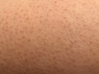
Keratosis pilaris is a common, autosomal-dominant, genetic condition of the skin's hair follicles characterized by the appearance of possibly itchy, small, gooseflesh-like bumps, with varying degrees of reddening or inflammation. It most often appears on the outer sides of the upper arms, thighs, face, back, and buttocks; KP can also occur on the hands, and tops of legs, sides, or any body part except glabrous (hairless) skin. Often the lesions can appear on the face, which may be mistaken for acne or folliculitis.

<span class="mw-page-title-main">Incontinentia pigmenti</span> Rare X-linked dominant genetic disorder

Incontinentia pigmenti (IP) is a rare X-linked dominant genetic disorder that affects the skin, hair, teeth, nails and central nervous system. It is named from its appearance under a microscope.

<span class="mw-page-title-main">Meleda disease</span> Medical condition

Meleda disease (MDM) or "mal de Meleda", also called Mljet disease, keratosis palmoplantaris and transgradiens of Siemens, is an extremely rare autosomal recessive congenital skin disorder in which dry, thick patches of skin develop on the soles of the hands and feet, a condition known as palmoplantar hyperkeratosis. Meleda Disease is a skin condition which usually can be identified not long after birth. This is a genetic condition but it is very rare. The hands and feet usually are the first to show signs of the disease but the disease can advance to other parts of the body. Signs of the disease include thickening of the skin, on hands and soles of feet, which can turn red in color. There currently is no cure and treatment is limited, but Acitretin can be used in severe cases.

<span class="mw-page-title-main">Urticaria pigmentosa</span> Most common form of cutaneous mastocytosis

Urticaria pigmentosa (also known as generalized eruption of cutaneous mastocytosis (childhood type) ) is the most common form of cutaneous mastocytosis. It is a rare disease caused by excessive numbers of mast cells in the skin that produce hives or lesions on the skin when irritated.

<span class="mw-page-title-main">Pseudoxanthoma elasticum</span> Medical condition

Pseudoxanthoma elasticum (PXE) is a genetic disease that causes mineralization of elastic fibers in some tissues. The most common problems arise in the skin and eyes, and later in blood vessels in the form of premature atherosclerosis. PXE is caused by autosomal recessive mutations in the ABCC6 gene on the short arm of chromosome 16 (16p13.1).

<span class="mw-page-title-main">Pruritic urticarial papules and plaques of pregnancy</span> Chronic rash that occurs during pregnancy

Pruritic urticarial papules and plaques of pregnancy (PUPPP), known in United Kingdom as polymorphic eruption of pregnancy (PEP), is a chronic hives-like rash that strikes some women during pregnancy. Some skin changes are known to occur in people who are pregnant while other skin conditions, or dermatoses, that people have prior to getting pregnant will become altered or symptoms will increase. Pruritic urticarial papules and plaques of pregnancy (PUPPP) is one of many skin conditions that is specific to pregnancy and occurs in about 1 in every 160 (0.625%) of pregnancies.

Conradi–Hünermann syndrome is a rare type of chondrodysplasia punctata. It is associated with the EBP gene and affects between one in 100,000 and one in 200,000 babies.

ATP2A2 also known as sarcoplasmic/endoplasmic reticulum calcium ATPase 2 (SERCA2) is an ATPase associated with Darier's disease and Acrokeratosis verruciformis.

<span class="mw-page-title-main">Netherton syndrome</span> Medical condition

Netherton syndrome is a severe, autosomal recessive form of ichthyosis associated with mutations in the SPINK5 gene. It is named after Earl W. Netherton (1910–1985), an American dermatologist who discovered it in 1958.

<span class="mw-page-title-main">Buschke–Ollendorff syndrome</span> Medical condition

Buschke–Ollendorff syndrome (BOS) is a rare genetic skin disorder associated with LEMD3, that typically presents with widespread painless papules.

<span class="mw-page-title-main">Urbach–Wiethe disease</span> Rare recessive genetic disorder

Urbach–Wiethe disease is a very rare recessive genetic disorder, with approximately 400 reported cases since its discovery. It was first officially reported in 1929 by Erich Urbach and Camillo Wiethe, although cases may be recognized dating back as early as 1908.

Kyrle disease is identified as a form of an acquired perforating disease. Other major perforating diseases are elastosis perforans serpiginosa and reactive perforating collagenosis. Recently, however, there is a controversy on categorizing Kyrle disease with perforating dermatosis or a subtype of acquired perforating collagenosis.

<span class="mw-page-title-main">Warty dyskeratoma</span> Benign tumour

Warty dyskeratoma, also known as an Isolated dyskeratosis follicularis, is a benign epidermal proliferation with distinctive histologic findings that may mimic invasive squamous cell carcinoma and commonly manifests as an umbilicated lesion with a keratotic plug, WD have some histopathologic similarities to viral warts but it's not caused by HPV and the majority of these lesions display overall histopathologic features consistent with a follicular adnexal neoplasm. Usually limited to the head, neck, scalp or face and vulva. Lesions are generally solitary and sporadic and may be associated with a follicular unit. Oral involvement, particularly the hard palate, and genital involvement have been reported. it can also be thought of as one of the manifestations of focal acantholytic dyskeratosis, an epidermal reaction pattern that can be seen in several disorders, including Darier's disease and Grover's disease. But the main Difference between Darier disease and Warty dyskeratoma, is that Darier disease inherited dermatosis consisting of multiple keratotic papules on the face, trunk, and extremities, while WD occurs as an isolated, noninherited, single keratotic nodule mainly confined to the head and neck as mentioned earlier.

Acrokeratosis verruciformis is a rare autosomal dominant disorder appearing at birth or in early childhood, characterized by skin lesions that are small, verrucous, flat papules resembling warts along with palmoplantar punctate keratoses and pits. However sporadic forms, whose less than 10 cases have been reported, presents at a later age, usually after the first decade and generally lack palmoplantar keratoses. Whether acrokeratosis verruciformis and Darier disease are related or distinct entities has been controversial, like Darier's disease, it is associated with defects in the ATP2A2 gene. however the specific mutations found in the ATP2A2 gene in acrokeratosis verruciformis have never been found in Darier's disease.

<span class="mw-page-title-main">Photoaging</span> Ultraviolet light-induced biological development over time

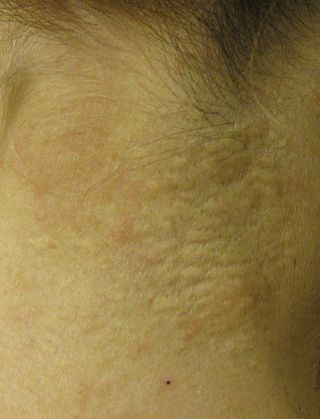
Photoaging or photoageing is a term used for the characteristic changes to skin induced by chronic UVA and UVB exposure. Tretinoin is the best studied retinoid in the treatment of photoaging.

Ichthyosis prematurity syndrome (IPS) is a dermatological disease with known genetic causes. This syndrome is a rare subcategory of autosomal recessive congenital ichthyosis (ARCI). It is associated with complications in the mid-trimester of a pregnancy leading to premature births. Although most prevalent in individuals of Scandinavian origin, there have also been scattered cases in people of Japanese, Italian and Indian ethnicity. This disorder is also referred to as ichthyosis congenital type IV.

<span class="mw-page-title-main">Familial disseminated comedones without dyskeratosis</span> Skin disorder from a rare gene mutation

Familial disseminated comedones without dyskeratosis (FDCWD) is a rare autosomal dominant skin disorder characterized by the presence of numerous comedones on the face, trunk, and extremities. The comedones are typically asymptomatic and do not lead to scarring. The disorder is thought to be caused by a mutation in the gene encoding the protein involucrin.

References

Notes

  1. 1 2 Rapini, Ronald P.; Bolognia, Jean L.; Jorizzo, Joseph L. (2007). Dermatology: 2-Volume Set. St. Louis, MO: Mosby. ISBN   978-1-4160-2999-1.
  2. Freedberg, et al. (2003). Fitzpatrick's Dermatology in General Medicine (6th ed.). McGraw-Hill. ISBN   0-07-138076-0.
  3. James, William; Berger, Timothy; Elston, Dirk (2005). Andrews' Diseases of the Skin: Clinical Dermatology (10th ed.). Saunders. ISBN   0-7216-2921-0.
  4. James, William D.; Elston, Dirk; Treat, James R.; Rosenbach, Misha A.; Neuhaus, Isaac (2020). "27. Genodermatoses and congenital anomalies". Andrews' Diseases of the Skin: Clinical Dermatology (13th ed.). Elsevier. p. 572. ISBN   978-0-323-54753-6.
  5. National Organization for Rare Disorders (2002). NORD Guide to Rare Disorders. Lippincott Williams & Wilkins. ISBN   0-7817-3063-5.
  6. Sehgal, V. N.; Srivastava, G. (2005). "Darier's (Darier-White) disease/keratosis follicularis". International Journal of Dermatology. 44 (3): 184–192. doi:10.1111/j.1365-4632.2004.02408.x. PMID   15807723. S2CID   45303870.
  7. "Darier disease | Genetic and Rare Diseases Information Center (GARD) – an NCATS Program". rarediseases.info.nih.gov. Retrieved 2020-05-08.
  8. 1 2 3 4 "Darier disease NZ". DermNet. Retrieved 2020-05-07.
  9. "Acrokeratosis Verruciformis of Hopf: Background, Pathophysiology, Etiology". 2020-03-26.{{cite journal}}: Cite journal requires |journal= (help)
  10. Monk, Sarah; Sakuntabhai, Anavaj; Carter, Simon A.; Bryce, Steven D.; Cox, Roger; Harrington, Louise; Levy, Elaine; Ruiz-Perez, Victor L.; Katsantoni, Eleni; Kodvawala, Ahmer; Munro, Colin S. (April 1998). "Refined Genetic Mapping of the Darier Locus to a <1-cM Region of Chromosome 12q24.1, and Construction of a Complete, High-Resolution P1 Artificial Chromosome/Bacterial Artificial Chromosome Contig of the Critical Region". The American Journal of Human Genetics. 62 (4): 890–903. doi:10.1086/301794. ISSN   0002-9297. PMC   1377034 . PMID   9529352.
  11. 1 2 Foggia, Lucie; Hovnanian, Alain (2004). "Calcium pump disorders of the skin". American Journal of Medical Genetics Part C: Seminars in Medical Genetics (in French). 131C (1): 20–31. doi: 10.1002/ajmg.c.30031 . ISSN   1552-4876. PMID   15468148. S2CID   675895.
  12. 1 2 3 Reference, Genetics Home. "Darier disease". Genetics Home Reference. Retrieved 2020-05-08.
  13. Sakuntabhai, Anavaj; Ruiz-Perez, Victor; Carter, Simon; Jacobsen, Nick; Burge, Susan; Monk, Sarah; Smith, Melanie; Munro, Colin S.; O'Donovan, Michael; Craddock, Nick; Kucherlapati, Raju (March 1999). "Mutations in ATP2A2, encoding a Ca 2+ pump, cause Darier disease". Nature Genetics. 21 (3): 271–277. doi:10.1038/6784. ISSN   1546-1718. PMID   10080178. S2CID   38684482.
  14. Ruiz-Perez, Victor L.; Carter, Simon A.; Healy, Eugene; Todd, Carole; Rees, Jonathan L.; Steijlen, Peter M.; Carmichael, Andrew J.; Lewis, Helen M.; Hohl, D.; Itin, Peter; Vahlquist, Anders (1999-09-01). "ATP2A2 Mutations in Darier's Disease: Variant Cutaneous Phenotypes Are Associated with Missense Mutations, But Neuropsychiatry Features Are Independent of Mutation Class". Human Molecular Genetics. 8 (9): 1621–1630. doi: 10.1093/hmg/8.9.1621 . ISSN   0964-6906. PMID   10441324.
  15. Sakuntabhai, Anavaj; Dhitavat, Jittima; Hovnanian, Alain; Burge, Susan (December 2000). "Mosaicism for ATP2A2 Mutations Causes Segmental Darier's Disease". Journal of Investigative Dermatology. 115 (6): 1144–1147. doi: 10.1046/j.1523-1747.2000.00182.x . ISSN   0022-202X. PMID   11121153.
  16. Gordon-Smith K, Jones LA, Burge SM, Munro CS, Tavadia S, Craddock N (September 2010). "The neuropsychiatric phenotype in Darier disease". Br. J. Dermatol. 163 (3): 515–22. doi:10.1111/j.1365-2133.2010.09834.x. PMID   20456342. S2CID   22856369.
  17. Hohl, D. Hand, J. Darier disease. (2017) Corona, R. (Ed) Uptodate.
  18. Lippincott's Illustrated Reviews: Biochemistry (Champe, Harvey & Ferrier, ISBN   0-7817-2265-9, 3rd ed., Lippincott Williams & Wilkins 2005)
  19. Kamijo M, Nishiyama C, Takagi A, Nakano N, Hara M, Ikeda S, Okumura K, Ogawa H (May 2012). "Cyclooxygenase-2 inhibition restores ultraviolet B-induced downregulation of ATP2A2/SERCA2 in keratinocytes: possible therapeutic approach of cyclooxygenase-2 inhibition for treatment of Darier disease". Br. J. Dermatol. 166 (5): 1017–22. doi:10.1111/j.1365-2133.2011.10789.x. PMID   22413864. S2CID   42918432.
  20. Millán-Parrilla F, Rodrigo-Nicolás B, Molés-Poveda P, Armengot-Carbó M, Quecedo-Estébanez E, Gimeno-Carpio E (April 2014). "Improvement of Darier disease with diclofenac sodium 3% gel". J. Am. Acad. Dermatol. 70 (4): e89–e90. doi: 10.1016/j.jaad.2013.11.033 . PMID   24629373.
  21. "Dermabrasion: MedlinePlus Medical Encyclopedia". medlineplus.gov. Retrieved 2020-05-08.
  22. "How do retinol creams work, anyway?". Sundaily. 30 November 2018. Retrieved 2020-05-08.
  23. Andrew's Diseases of the Skin (James, Berger, Elston, 10th ed., Saunders Elsevier, 2006)
  24. "FIRST Skin Foundation". Archived from the original on 2019-05-30. Retrieved 2020-05-08.
  25. Godic A, Miljkovic J, Kansky A, Vidmar G (June 2005). "Epidemiology of Darier's Disease in Slovenia". Acta Dermatovenerol Alp Pannonica Adriat. 14 (2): 43–8. PMID   16001099.
  26. Munro CS (August 1992). "The phenotype of Darier's disease: penetrance and expressivity in adults and children". Br. J. Dermatol. 127 (2): 126–30. doi:10.1111/j.1365-2133.1992.tb08044.x. PMID   1390140. S2CID   2911858.
  27. Tavadia S, Mortimer E, Munro CS (January 2002). "Genetic epidemiology of Darier's disease: a population study in the west of Scotland". Br. J. Dermatol. 146 (1): 107–9. doi:10.1046/j.1365-2133.2002.04559.x. PMID   11841374. S2CID   42621572.
  28. Burge SM, Wilkinson JD (July 1992). "Darier-White disease: a review of the clinical features in 163 patients". J. Am. Acad. Dermatol. 27 (1): 40–50. doi:10.1016/0190-9622(92)70154-8. PMID   1619075.
  29. Crissey, John Thorne; Parish, Lawrence C.; Holubar, Karl (2013). "Late nineteenth century French dermatology". Historical Atlas of Dermatology and Dermatologists. CRC Press. p. 75. ISBN   978-1-84184-864-8.
  30. Cadogan, Dr Mike (2019-03-01). "Ferdinand-Jean Darier • LITFL". Life in the Fast Lane • LITFL • Medical Blog. Retrieved 2020-05-08.
  31. "Keratosis Follicularis (Darier Disease): Background, Pathophysiology, Epidemiology". Medscape . 2020-10-01.
  32. "Was he murdered?". The New Paper. 29 December 1995.
  33. "Chicken slaughterer charged with murder". The Straits Times. 4 January 1996.
  34. "Man gets death for murdering opium addict, 82". The Straits Times. 13 August 1996.
  35. "Did man's skin disease lead him to kill addict?". The Straits Times. 24 February 1998.
  36. "Skin disease may save man's life". The Straits Times. 18 April 1998.
  37. "Skin disease saves man's neck". The Straits Times. 28 May 1998.

Sources