Related Research Articles
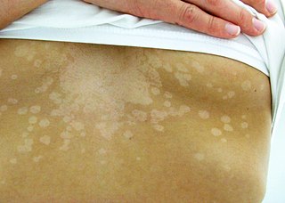
Tinea versicolor is a condition characterized by a skin eruption on the trunk and proximal extremities. The majority of tinea versicolor is caused by the fungus Malassezia globosa, although Malassezia furfur is responsible for a small number of cases. These yeasts are normally found on the human skin and become troublesome only under certain conditions, such as a warm and humid environment, although the exact conditions that cause initiation of the disease process are poorly understood.

Malassezia is a genus of fungi. It is the sole genus in family Malasseziaceae, which is the only family in order Malasseziales, itself the single member of class Malasseziomycetes. Malassezia species are naturally found on the skin surfaces of many animals, including humans. In occasional opportunistic infections, some species can cause hypopigmentation or hyperpigmentation on the trunk and other locations in humans. Allergy tests for these fungi are available. It is believed French revolutionary Jean-Paul Marat suffered from a fungal infection from Malassezia restricta, which lead to his frequent bathing in a medicinal substance.

Chromoblastomycosis is a long-term fungal infection of the skin and subcutaneous tissue.

Lobomycosis is a fungal infection of the skin. It usually presents with bumps in the skin, firm swellings, deep skin lesions, or malignant tumors.

Pythiosis is a rare and deadly tropical disease caused by the oomycete Pythium insidiosum. Long regarded as being caused by a fungus, the causative agent was not discovered until 1987. It occurs most commonly in horses, dogs, and humans, with isolated cases in other large mammals. The disease is contracted after exposure to stagnant fresh water such as swamps, ponds, lakes, and rice paddies. P. insidiosum is different from other members of the genus in that human and horse hair, skin, and decaying animal and plant tissue are chemoattractants for its zoospores. Additionally, it is the only member in the genus known to infect mammals, while other members are pathogenic to plants and are responsible for some well-known plant diseases.

Paracoccidioidomycosis (PCM), also known as South American blastomycosis, is a fungal infection that can occur as a mouth and skin type, lymphangitic type, multi-organ involvement type (particularly lungs), or mixed type. If there are mouth ulcers or skin lesions, the disease is likely to be widespread. There may be no symptoms, or it may present with fever, sepsis, weight loss, large glands, or a large liver and spleen.

Paracoccidioides is a genus of fungi in the order Onygenales. Species are known human pathogens producing yeast-like states under pathogenic conditions. They include the causative agents of paracoccidioidomycosis and lobomycosis.
Otomycosis is a fungal ear infection, a superficial mycotic infection of the outer ear canal caused by micro-organisms called fungi which are related to yeast and mushrooms. It is more common in tropical or warm countries. The infection may be either subacute or acute and is characterized by itching in the ear, malodorous discharge, inflammation, pruritus, scaling, and severe discomfort or ear pain. The mycosis results in inflammation, superficial epithelial exfoliation, masses of debris containing hyphae, suppuration, and pain. Otomycosis can also cause hearing loss.

Sporothrix schenckii, a fungus that can be found worldwide in the environment, is named for medical student Benjamin Schenck, who in 1896 was the first to isolate it from a human specimen. The species is present in soil as well as in and on living and decomposing plant material such as peat moss. It can infect humans as well as animals and is the causative agent of sporotrichosis, commonly known as "rose handler's disease." The most common route of infection is the introduction of spores to the body through a cut or puncture wound in the skin. Infection commonly occurs in otherwise healthy individuals but is rarely life-threatening and can be treated with antifungals. In the environment it is found growing as filamentous hyphae. In host tissue it is found as a yeast. The transition between the hyphal and yeast forms is temperature dependent making S. schenckii a thermally dimorphic fungus.

Paracoccidioides brasiliensis is a dimorphic fungus and one of the two species that cause paracoccidioidomycosis. The fungus has been affiliated with the family Ajellomycetaceae although a sexual state or teleomorph has not yet been found.

Blastomyces dermatitidis is a dimorphic fungus that causes blastomycosis, an invasive and often serious fungal infection found occasionally in humans and other animals. It lives in soil and wet, decaying wood, often in an area close to a waterway such as a lake, river or stream. Indoor growth may also occur, for example, in accumulated debris in damp sheds or shacks. The fungus is endemic to parts of eastern North America, particularly boreal northern Ontario, southeastern Manitoba, Quebec south of the St. Lawrence River, parts of the U.S. Appalachian mountains and interconnected eastern mountain chains, the west bank of Lake Michigan, the state of Wisconsin, and the entire Mississippi Valley including the valleys of some major tributaries such as the Ohio River. In addition, it occurs rarely in Africa both north and south of the Sahara Desert, as well as in the Arabian Peninsula and the Indian subcontinent. Though it has never been directly observed growing in nature, it is thought to grow there as a cottony white mold, similar to the growth seen in artificial culture at 25 °C (77 °F). In an infected human or animal, however, it converts in growth form and becomes a large-celled budding yeast. Blastomycosis is generally readily treatable with systemic antifungal drugs once it is correctly diagnosed; however, delayed diagnosis is very common except in highly endemic areas.

Dimorphic fungi are fungi that can exist in the form of both mold and yeast. This is usually brought about by change in temperature and the fungi are also described as thermally dimorphic fungi. An example is Talaromyces marneffei, a human pathogen that grows as a mold at room temperature, and as a yeast at human body temperature.

Fonsecaea pedrosoi is a fungal species in the family Herpotrichiellaceae, and the major causative agent of chromoblastomycosis. This species is commonly found in tropical and sub-tropical regions, especially in South America, where it grows as a soil saprotroph. Farming activities in the endemic zone are a risk factor for the development of chromoblastomycosis.
Phaeohyphomycosis is a diverse group of fungal infections, caused by dematiaceous fungi whose morphologic characteristics in tissue include hyphae, yeast-like cells, or a combination of these. It can be associated with an array of melanistic filamentous fungi including Alternaria species, Exophiala jeanselmei, and Rhinocladiella mackenziei.
Histoplasma duboisii is a saprotrophic fungus responsible for the invasive infection known as African histoplasmosis. This species is a close relative of Histoplasma capsulatum, the agent of classical histoplasmosis, and the two occur in similar habitats. Histoplasma duboisii is restricted to continental Africa and Madagascar, although scattered reports have arisen from other places usually in individuals with an African travel history. Like, H. capsulatum, H. duboisii is dimorphic – growing as a filamentous fungus at ambient temperature and a yeast at body temperature. It differs morphologically from H. capsulatum by the typical production of a large-celled yeast form. Both agents cause similar forms of disease, although H. duboisii predominantly causes cutaneous and subcutaneous disease in humans and non-human primates. The agent responds to many antifungal drug therapies used to treat serious fungal diseases.
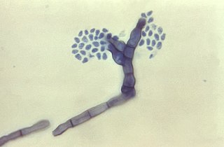
Fonsecaea compacta is a saprophytic fungal species found in the family Herpotrichiellaceae. It is a rare etiological agent of chromoblastomycosis, with low rates of correspondence observed from reports. The main active components of F. compacta are glycolipids, yet very little is known about its composition. F. compacta is widely regarded as a dysplastic variety of Fonsecaea pedrosoi, its morphological precursor. The genus Fonsecaea presently contains two species, F. pedrosoi and F. compacta. Over 100 strains of F. pedrosoi have been isolated but only two of F. compacta.

Trichosporon asteroides is an asexual basidiomycetous fungus first described from human skin but now mainly isolated from blood and urine. T. asteroides is a hyphal fungus with a characteristically yeast-like appearance due to the presence of slimy arthroconidia. Infections by this species usually respond to treatment with azoles and amphotericin B.
Arthrographis kalrae is an ascomycetous fungus responsible for human nail infections described in 1938 by Cochet as A. langeronii. A. kalrae is considered a weak pathogen of animals including human restricted to the outermost keratinized layers of tissue. Infections caused by this species are normally responsive to commonly used antifungal drugs with only very rare exceptions.

Paracoccidioides lutzii is a dimorphic fungus that is one of the causal agents of paracoccidioidomycosis, together with Paracoccidioides brasiliensis. Unlike P. brasiliensis, which is found throughout Central and South America, P. lutzii is found only in Brazil and Ecuador. It is less virulent than P. brasiliensis.

Libero Ajello was an American mycologist. He cofounded and was first president of the International Society of Human and Animal Mycology (ISHAM). He was the head of the Division of Mycotic Diseases at the Communicable Disease Center (CDC), and editor of the ISHAM Journal Medical Mycology for several years. He was one of the first researchers to investigate histoplasmosis and coccidioidomycosis in the United States and made valuable contributions to the comprehensive field of veterinary and human fungal disease diagnosis.
References
- ↑ Herr RA, Tarcha EJ, Taborda PR, Taylor JW, Ajello L, Mendoza L (2001). "Phylogenetic analysis of Lacazia loboi places this previously uncharacterized pathogen within the dimorphic Onygenales". J. Clin. Microbiol. 39 (1): 309–14. doi:10.1128/JCM.39.1.309-314.2001. PMC 87720 . PMID 11136789.
- ↑ Taborda, P. R., V. A. Taborda, and M. R. McGinnis. 1999. Lacazia loboi gen. nov., comb, nov., the etiologic agent of lobomycosis. J Clin Microbiol. 37:2031-2033.
- ↑ Burns, R. A., J. S. Roy, C. Woods, A. A. Padhye, and D. W. Warnock. 2000. Report of the first human case of lobomycosis in the United States. J Clin Microbiol. 38:1283-5.
- 1 2 3 4 5 Collier, L., A. Balows, and M. Sussman. 1998. Topley & Wilson's Microbiology and Microbial Infections, 9th ed, vol. 4. Arnold, London, Sydney, Auckland, New York.
- ↑ Jaramillo, D., A. Cortes, A. Restrepo, M. Builes, and M. Robledo. 1976. Lobomycosis. Report of the eighth Colombian case and review of the literature. J Cutan Pathol. 3:180-9.
- ↑ Rodriguez-Toro, G. 1993. Lobomycosis. Int. J. Dermatol. 32:324-32.
- ↑ Rodriguez-Toro, G., and N. Tellez. 1992. Lobomycosis in Colombian Amer Indian patients. Mycopathologia. 120:5-9.
- ↑ Haubold, E. M., J. F. Aronson, D. F. Cowan, M. R. McGinnis, and C. R. Cooper. 1998. Isolation of fungal rDNA from bottlenose dolphin skin infected with Loboa loboi. Med Mycol. 36:263-267.
- ↑ Camargo, Z. P., R. G. Baruzzi, S. M. Maeda, and M. C. Floriano. 1998. Antigenic relationship between Loboa loboi and Paracoccidioides brasiliensis as shown by serological methods. Med Mycol. 36:413-417.
- ↑ Restrepo, A. 1994. Treatment of tropical mycoses. J. Amer. Acad. Dermatol. 31:S91-102.
- ↑ Mendoza L, Vilela R, Rosa PS, Fernandes Belone AF (December 2005). "Lacazia loboi and Rhinosporidium seeberi: a genomic perspective". Rev Iberoam Micol. 22 (4): 213–6. doi:10.1016/S1130-1406(05)70045-0. PMID 16499413.