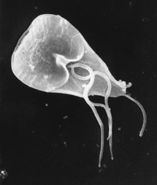
Giardia duodenalis, also known as Giardia intestinalis and Giardia lamblia, is a flagellated parasitic microorganism of the genus Giardia that colonizes the small intestine, causing a diarrheal condition known as giardiasis. The parasite attaches to the epithelium by a ventral adhesive disc or sucker, and reproduces via binary fission. Giardiasis does not spread via the bloodstream, nor does it spread to other parts of the gastrointestinal tract, but remains confined to the lumen of the small intestine. Giardia has an outer membrane that makes it possible to retain life, even when outside of the host body, and which can make it tolerant to chlorine disinfection. Giardia trophozoites absorb their nutrients from the lumen, and are anaerobes. If the organism is split and stained, its characteristic pattern resembles the familiar "smiley face" symbol.

Coccidia (Coccidiasina) are a subclass of microscopic, spore-forming, single-celled obligate intracellular parasites belonging to the apicomplexan class Conoidasida. As obligate intracellular parasites, they must live and reproduce within an animal cell. Coccidian parasites infect the intestinal tracts of animals, and are the largest group of apicomplexan protozoa.

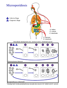
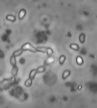
Microsporidia are a group of spore-forming unicellular parasites. These spores contain an extrusion apparatus that has a coiled polar tube ending in an anchoring disc at the apical part of the spore. They were once considered protozoans or protists, but are now known to be fungi, or a sister group to fungi. These fungal microbes are obligate eukaryotic parasites that use a unique mechanism to infect host cells. They have recently been discovered in a 2017 Cornell study to infect Coleoptera on a large scale. So far, about 1500 of the probably more than one million species are named. Microsporidia are restricted to animal hosts, and all major groups of animals host microsporidia. Most infect insects, but they are also responsible for common diseases of crustaceans and fish. The named species of microsporidia usually infect one host species or a group of closely related taxa. Approximately 10 percent of the species are parasites of vertebrates —several species, most of which are opportunistic, can infect humans, in whom they can cause microsporidiosis.

Fumagillin is a complex biomolecule and used as an antimicrobial agent. It was isolated in 1949 from the microbial organism Aspergillus fumigatus.

An opportunistic infection is an infection caused by pathogens that take advantage of an opportunity not normally available. These opportunities can stem from a variety of sources, such as a weakened immune system, an altered microbiome, or breached integumentary barriers. Many of these pathogens do not necessarily cause disease in a healthy host that has a non-compromised immune system, and can, in some cases, act as commensals until the balance of the immune system is disrupted. Opportunistic infections can also be attributed to pathogens which cause mild illness in healthy individuals but lead to more serious illness when given the opportunity to take advantage of an immunocompromised host.

Cutaneous larva migrans is a skin disease in humans, caused by the larvae of various nematode parasites of the hookworm family (Ancylostomatidae). The parasites live in the intestines of dogs, cats, and wild animals; they should not be confused with other members of the hookworm family for which humans are definitive hosts, namely Ancylostoma duodenale and Necator americanus.

Retortamonas is a genus of flagellated excavates. It is one of only two genera belonging to the family Retortamonadidae along with the genus Chilomastix. The genus parasitizes a large range of hosts including humans. Species within this genus are considered harmless commensals which reside in the intestine of their host. The wide host diversity is a useful factor given that species are distinguished based on their host rather than morphology. This is because all species share similar morphology, which would present challenges when trying to make classifications based on structural anatomy. Although Retortamonas currently includes over 25 known species, it is possible that some defined species are synonymous, given that such overlapping species have been discovered in the past. Further efforts into learning about this genus must be done such as cross-transmission testing as well as biochemical and genetic studies. One of the most well-known species within this genus is Retortamonas intestinalis, a human parasite that lives in the large intestine of humans.
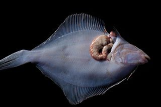
A xenoma is a growth caused by various protists and fungi, most notably microsporidia. It can occur on numerous organisms; however is predominantly found on fish.
Enterocytozoon bieneusi is a species of the order Chytridiopsida which infects the intestinal epithelial cells. It is an obligate intracellular parasite.
Antiparasitics are a class of medications which are indicated for the treatment of parasitic diseases, such as those caused by helminths, amoeba, ectoparasites, parasitic fungi, and protozoa, among others. Antiparasitics target the parasitic agents of the infections by destroying them or inhibiting their growth; they are usually effective against a limited number of parasites within a particular class. Antiparasitics are one of the antimicrobial drugs which include antibiotics that target bacteria, and antifungals that target fungi. They may be administered orally, intravenously or topically. Overuse or misuse of antiparasitics can lead to the development of antimicrobial resistance.
Encephalitozoon intestinalis is a parasite. It can cause microsporidiosis.
Karyolysus is a genus of coccidia. With the exception of K. sonomae whose vertebrate host is the yellow-legged frog, species in this genus only infect lizards of the genus Lacerta.

Encephalitozoon cuniculi is a microsporidial pathogen of mammals with world-wide distribution. An important cause of neurologic and renal disease in rabbits, E. cuniculi can also cause disease in immunocompromised people.
Acroeimeria is a genus of parasites that contains those species which initially develop immediately beneath the brush-border of the intestinal epithelium, but the meronts and gamonts of which are early on extruded to form a layer on the surface of the gut mucosa. Morphologically they are similar to the Eimeria to which they are closely related. The genus was described in 1989 by Paperna and Landsberg.

Cystoisospora belli, previously known as Isospora belli, is a parasite that causes an intestinal disease known as cystoisosporiasis. This protozoan parasite is opportunistic in immune suppressed human hosts. It primarily exists in the epithelial cells of the small intestine, and develops in the cell cytoplasm. The distribution of this coccidian parasite is cosmopolitan, but is mainly found in tropical and subtropical areas of the world such as the Caribbean, Central and S. America, India, Africa, and S.E. Asia. In the U.S., it is usually associated with HIV infection and institutional living.

The parasitophorous vacuole (PV) is a structure produced by apicomplexan parasites in the cells of its host. The PV allows the parasite to develop while protected from the phagolysosomes of the host cell.
Ordospora colligata is an intracellular parasite belonging to the Microsporidia. It is an obligatory gut parasite with the crustacean Daphnia magna as its only host. So far it has been reported from Europe and Asia.
Nematocida parisii is a parasitic species of Microsporidia fungi found in wild isolates of the common nematode, Caenorhabditis elegans. The fungus forms spores and replicates in the intestines before leaving the host.
Mycotypha microspora, also known as Microtypha microspora, is a filamentous fungus in the division Zygomycota. It was discovered in a Citrus aurantium peel in 1932 by E. Aline Fenner, who proposed a new genus Mycotypha to accommodate it. Mycotypha africana, which is another species in the genus Mycotypha, is closely related to M. microspora. The fungus has subsequently been isolated from both outdoor and indoor settings around the world, and is typically found in soil and dung. The species rarely causes infections in humans, but has recently been involved in the clinical manifestation of the life-threatening disease mucormycosis.

Enterospora nucleophila is a microsporidian infecting the gilt-head bream. It develops primarily within the nuclei of rodlet cells and enterocytes, at the intestinal epithelium. It can also be found in cytoplasmic position within other cell types, including phagocytes, at subepithelial layers. It is the causative agent of emaciative microsporidiosis of gilthead sea bream, a chronic condition manifested as a severe growth arrestment, normally accompanied by trickling mortality.