Sporotrichosis

Last updated
Sporotrichosis
Other namesRose thorn disease, rose gardener's disease, [1] rose handler's disease [2]
Feline sporotrichosis 4.jpg
Cytologic preparation from a case of feline sporotrichosis; phagocytic cells show numerous variably-shaped yeast forms within
Specialty Infectious disease
Symptoms Firm painless nodules that later ulcerate. [3]
Causes Sporothrix schenckii [1]
Diagnostic method
  • Serology [1]
  • Culture: Skin, sputum, CSF, joint aspirate [1]
Differential diagnosis Leishmaniasis, nocardiosis, mycobacterium marinum, [3] cat-scratch disease, syphilis, leprosy, sarcoidosis, tuberculosis [1]
Treatment Antifungals, surgery [1]
Medication Itraconazole, posaconazole, amphotericin B [1]
Prognosis Good with treatment, poor if widespread disease [1]

Sporotrichosis, also known as rose handler's disease, [2] is a fungal infection that may be localised to skin, lungs, bone and joint, or become systemic. [2] [4] It presents with firm painless nodules that later ulcerate. [3] Following initial exposure to Sporothrix schenckii , the disease typically progresses over a period of a week to several months. [1] Serious complications may develop in people who have a weakened immune system. [1]

Contents

Sporotrichosis is caused by fungi of the S. schenckii species complex. [5] [6] Because S. schenckii is naturally found in soil, hay, sphagnum moss, and plants, it most often affects farmers, gardeners, and agricultural workers. [7] It enters through small cuts in the skin to cause a fungal infection. [1] In cases of sporotrichosis affecting the lungs, the fungal spores enter by inhalation. [1] Sporotrichosis can be acquired by handling cats with the disease; it is an occupational hazard for veterinarians. [1]

Treatment depends on the site and extent of infection. [1] Topical antifungals may be applied to skin lesions. [1] Deep infection in the lungs may require surgery. [1] Systemic medications used include Itraconazole, posaconazole and amphotericin B. [1] With treatment, most people will recover, [1] but an immunocompromised status and systemic infection carry a worse prognosis. [1]

S. schenkii, the causal fungus, is found worldwide. [1] The species was named for Benjamin Schenck, a medical student who, in 1896, was the first to isolate it from a human specimen. [8]

Sporotrichosis has been reported in cats, [1] mules, dogs, mice and rats. [3]

Signs and symptoms

Sporotrichosis by the fungus Sporothrix schenckii Sporotrichosis by the fungus Sporothrix schenckii PHIL 3940 lores.jpg
Sporotrichosis by the fungus Sporothrix schenckii
This is the most common form of this disease. Symptoms of this form include nodular lesions or bumps in the skin, at the point of entry and also along lymph nodes and vessels. The lesion starts off small and painless, and ranges in color from pink to purple. Left untreated, the lesion becomes larger and look similar to a boil and more lesions will appear, until a chronic ulcer develops.[ citation needed ]
Usually, cutaneous sporotrichosis lesions occur in the finger, hand, and arm.[ citation needed ]
This rare form of the disease occur when S. schenckii spores are inhaled. Symptoms of pulmonary sporotrichosis include productive coughing, nodules and cavitations of the lungs, fibrosis, and swollen hilar lymph nodes. Patients with this form of sporotrichosis are susceptible to developing tuberculosis and pneumonia [ citation needed ]
When the infection spreads from the initial site to secondary sites in the body, the disease develops into an uncommon and potentially life-threatening form, called disseminated sporotrichosis. The infection can spread to joints and bones (called osteoarticular sporotrichosis) as well as the central nervous system and the brain (called sporotrichosis meningitis).[ citation needed ]
Some symptoms of disseminated sporotrichosis include weight loss, anorexia, and bone lesions.

Complications

Open sporotrichosis lesions on the skin will on occasion become superinfected with bacteria. [9] Cellulitis may also occur. [9]

Diagnosis

Conidiophores and conidia of the fungus Sporothrix schenckii Conidiophores and conidia of the fungus Sporothrix schenckii PHIL 4208 lores.jpg
Conidiophores and conidia of the fungus Sporothrix schenckii

Sporotrichosis is an acute infection with slow progression and often subtle symptoms. It is often difficult to diagnose, as many other diseases share similar symptoms and therefore must be ruled out.[ citation needed ]

Patients with sporotrichosis will have antibodies against the fungus S. schenckii; however, due to variability in sensitivity and specificity, antibody detection may not be a reliable diagnostic test for this disease. The confirming diagnosis remains culturing the fungus from the skin, sputum, synovial fluid, and cerebrospinal fluid. Smears should be taken from any draining fistulas or ulcers.[ citation needed ]

Cats with sporotrichosis are unique in that the exudate from their lesions may contain numerous infectious organisms. This makes cytological evaluation of exudate a valuable diagnostic tool in this species. Exudate is pyogranulomatous, and phagocytic cells may be packed with yeast forms. These yeast cells are variable in size; many are cigar-shaped.[ citation needed ]

Differential diagnosis

Differential diagnoses includes: leishmaniasis, nocardiosis, mycobacterium marinum, [3] cat-scratch disease, leprosy, syphilis, sarcoidosis and tuberculosis. [1]

Prevention

The majority of sporotrichosis cases occur when the fungus is introduced through a cut or puncture in the skin while handling vegetation containing the fungal spores. Prevention of this disease includes wearing long sleeves and gloves while working with soil, hay bales, rose bushes, pine seedlings, and sphagnum moss.

The risk of sporotrichosis in cats is increased in male cats that roam outdoors. [10] Accordingly, the risk may be reduced by keeping cats indoors or neutering them. [11] Isolating infected animals can also be a preventive measure. [11] [12] The risk of spread from infected cats to humans can be reduced by appropriate biosafety measures, including wearing personal protective equipment when handling a cat with suspected sporotrichosis and by washing hands, arms and clothing after handling the cat. [12]

Treatment

Treatment of sporotrichosis depends on the severity and location of the disease. The following are treatment options for this condition: [13]

Potassium iodide is an anti-fungal drug that is widely used as a treatment for cutaneous sporotrichosis. Despite its wide use, there is no high-quality evidence for or against this practice. Further studies are needed to assess the efficacy and safety of oral potassium iodide in the treatment of sporotrichosis. [14]
These are antifungal drugs. Itraconazole is currently the drug of choice and is significantly more effective than fluconazole. Fluconazole should be reserved for patients who cannot tolerate itraconazole.
This antifungal medication is delivered intravenously. Many patients, however, cannot tolerate Amphotericin B due to its potential side effects of fever, nausea, and vomiting.
Lipid formulations of amphotericin B are usually recommended instead of amphotericin B deoxycholate because of a better adverse-effect profile. Amphotericin B can be used for severe infection during pregnancy. For children with disseminated or severe disease, amphotericin B deoxycholate can be used initially, followed by itraconazole. [15]
In case of sporotrichosis meningitis, the patient may be given a combination of Amphotericin B and 5-fluorocytosine/Flucytosine.
500mg and 1000mg daily dosages of terbinafine for twelve to 24 weeks has been used to treat cutaneous sporotrichosis. [16]
Several studies have shown that posaconazole has in vitro activity similar to that of amphotericin B and itraconazole; therefore, it shows promise as an alternative therapy. However, voriconazole susceptibility varies. Because the correlation between in vitro data and clinical response has not been demonstrated, there is insufficient evidence to recommend either posaconazole or voriconazole for treatment of sporotrichosis at this time. [15]
In cases of bone infection and cavitary nodules in the lungs, surgery may be necessary.
Heat creates higher tissue temperatures, which may inhibit fungus growth while the immune system counteracts the infection. The "pocket warmer" used for this purpose has the advantage of being able to maintain a constant temperature of 44 degrees-45 degrees C on the skin surface for several hours, while permitting unrestricted freedom of movement. The duration of treatment depends on the type of lesion, location, depth, and size. Generally, local application for 1-2 h per day, or in sleep time, for 5-6 weeks seems to be sufficient. [17]

Other animals

Ulcerative skin disease in a cat with sporotrichosis; a cat with this disease must be handled with caution as this form can be contagious to other animals and to humans Feline sporotrichosis 3.jpg
Ulcerative skin disease in a cat with sporotrichosis; a cat with this disease must be handled with caution as this form can be contagious to other animals and to humans

Sporotrichosis can be diagnosed in domestic and wild mammals. In veterinary medicine it is most frequently seen in cats and horses. Cats have a particularly severe form of cutaneous sporotrichosis. Infected cats may exhibit abscesses, cellulitis, or draining wounds that fail to respond to antibiotic treatment. [10]

Sporotrichosis can spread from nonhuman animals to humans (zoonosis). Infected cats in particular exude large quantities of Sporothrix organisms from their skin leasions and can spread the infection to people who handle them. [10] [18] Although cats are the most common animal source, the infection has also been known to spread to humans from dogs, rats, squirrels, and armadillos. [19]

See also

Related Research Articles

<span class="mw-page-title-main">Coccidioidomycosis</span> Fungal infection

Coccidioidomycosis, commonly known as cocci, Valley fever, as well as California fever, desert rheumatism, or San Joaquin Valley fever, is a mammalian fungal disease caused by Coccidioides immitis or Coccidioides posadasii. Coccidioidomycosis is endemic in certain parts of the United States in Arizona, California, Nevada, New Mexico, Texas, Utah, and northern Mexico.

<span class="mw-page-title-main">Histoplasmosis</span> Human disease

Histoplasmosis is a fungal infection caused by Histoplasma capsulatum. Symptoms of this infection vary greatly, but the disease affects primarily the lungs. Occasionally, other organs are affected; called disseminated histoplasmosis, it can be fatal if left untreated.

<span class="mw-page-title-main">Cryptococcosis</span> Potentially fatal fungal disease

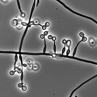
Cryptococcosis is a potentially fatal fungal infection of mainly the lungs, presenting as a pneumonia, and brain, where it appears as a meningitis. Cough, difficulty breathing, chest pain and fever are seen when the lungs are infected. When the brain is infected, symptoms include headache, fever, neck pain, nausea and vomiting, light sensitivity and confusion or changes in behavior. It can also affect other parts of the body including skin, where it may appear as several fluid-filled nodules with dead tissue.

<i>Coccidioides immitis</i> Species of fungus

Coccidioides immitis is a pathogenic fungus that resides in the soil in certain parts of the southwestern United States, northern Mexico, and a few other areas in the Western Hemisphere.

<span class="mw-page-title-main">Blastomycosis</span> Medical condition

Blastomycosis, also known as Gilchrist's disease, is a fungal infection, typically of the lungs, which can spread to brain, stomach, intestine and skin, where it appears as crusting purplish warty plaques with a roundish bumpy edge and central depression. Only about half of people with the disease have symptoms, which can include fever, cough, night sweats, muscle pains, weight loss, chest pain, and feeling tired. Symptoms usually develop between three weeks and three months after breathing in the spores. In 25% to 40% of cases, the infection also spreads to other parts of the body, such as the skin, bones or central nervous system. Although blastomycosis is especially dangerous for those with weak immune systems, most people diagnosed with blastomycosis have healthy immune systems.

<span class="mw-page-title-main">Tinea capitis</span> Cutaneous fungal infection of the scalp

Tinea capitis is a cutaneous fungal infection (dermatophytosis) of the scalp. The disease is primarily caused by dermatophytes in the genera Trichophyton and Microsporum that invade the hair shaft. The clinical presentation is typically single or multiple patches of hair loss, sometimes with a 'black dot' pattern, that may be accompanied by inflammation, scaling, pustules, and itching. Uncommon in adults, tinea capitis is predominantly seen in pre-pubertal children, more often boys than girls.

<span class="mw-page-title-main">Chromoblastomycosis</span> Medical condition

Chromoblastomycosis is a long-term fungal infection of the skin and subcutaneous tissue.

<span class="mw-page-title-main">Dermatophytosis</span> Fungal infection of the skin

Dermatophytosis, also known as ringworm, is a fungal infection of the skin. Typically it results in a red, itchy, scaly, circular rash. Hair loss may occur in the area affected. Symptoms begin four to fourteen days after exposure. Multiple areas can be affected at a given time.

<span class="mw-page-title-main">Onychomycosis</span> Medical condition

Onychomycosis, also known as tinea unguium, is a fungal infection of the nail. Symptoms may include white or yellow nail discoloration, thickening of the nail, and separation of the nail from the nail bed. Toenails or fingernails may be affected, but it is more common for toenails. Complications may include cellulitis of the lower leg. A number of different types of fungus can cause onychomycosis, including dermatophytes and Fusarium. Risk factors include athlete's foot, other nail diseases, exposure to someone with the condition, peripheral vascular disease, and poor immune function. The diagnosis is generally suspected based on the appearance and confirmed by laboratory testing.

<span class="mw-page-title-main">Pythiosis</span> Tropical disease caused by the oomycete Pythium insidiosum

Pythiosis is a rare and deadly tropical disease caused by the oomycete Pythium insidiosum. Long regarded as being caused by a fungus, the causative agent was not discovered until 1987. It occurs most commonly in horses, dogs, and humans, with isolated cases in other large mammals. The disease is contracted after exposure to stagnant fresh water such as swamps, ponds, lakes, and rice paddies. P. insidiosum is different from other members of the genus in that human and horse hair, skin, and decaying animal and plant tissue are chemoattractants for its zoospores. Additionally, it is the only member in the genus known to infect mammals, while other members are pathogenic to plants and are responsible for some well-known plant diseases.

Talaromycosis is a fungal infection that presents with painless skin lesions of the face and neck, as well as an associated fever, anaemia, and enlargement of the lymph glands and liver.

<span class="mw-page-title-main">Aspergillosis</span> Medical condition

Aspergillosis is a fungal infection of usually the lungs, caused by the genus Aspergillus, a common mould that is breathed in frequently from the air around, but does not usually affect most people. It generally occurs in people with lung diseases such as asthma, cystic fibrosis or tuberculosis, or those who have had a stem cell or organ transplant, and those who cannot fight infection because of medications they take such as steroids and some cancer treatments. Rarely, it can affect skin.

<span class="mw-page-title-main">Paracoccidioidomycosis</span> Medical condition

Paracoccidioidomycosis (PCM), also known as South American blastomycosis, is a fungal infection that can occur as a mouth and skin type, lymphangitic type, multi-organ involvement type (particularly lungs), or mixed type. If there are mouth ulcers or skin lesions, the disease is likely to be widespread. There may be no symptoms, or it may present with fever, sepsis, weight loss, large glands, or a large liver and spleen.

<i>Sporothrix schenckii</i> Species of fungus

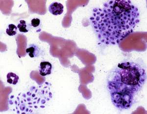
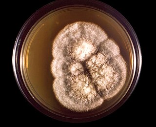
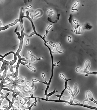
Sporothrix schenckii, a fungus that can be found worldwide in the environment, is named for medical student Benjamin Schenck, who in 1896 was the first to isolate it from a human specimen. The species is present in soil as well as in and on living and decomposing plant material such as peat moss. It can infect humans as well as animals and is the causative agent of sporotrichosis, commonly known as "rose handler's disease." The most common route of infection is the introduction of spores to the body through a cut or puncture wound in the skin. Infection commonly occurs in otherwise healthy individuals but is rarely life-threatening and can be treated with antifungals. In the environment it is found growing as filamentous hyphae. In host tissue it is found as a yeast. The transition between the hyphal and yeast forms is temperature dependent making S. schenckii a thermally dimorphic fungus.

<i>Ochroconis gallopava</i> Species of fungus

Ochroconis gallopava, also called Dactylaria gallopava or Dactylaria constricta var. gallopava, is a member of genus Dactylaria. Ochroconis gallopava is a thermotolerant, darkly pigmented fungus that causes various infections in fowls, turkeys, poults, and immunocompromised humans first reported in 1986. Since then, the fungus has been increasingly reported as an agent of human disease especially in recipients of solid organ transplants. Ochroconis gallopava infection has a long onset and can involve a variety of body sites. Treatment of infection often involves a combination of antifungal drug therapy and surgical excision.

<i>Phialophora verrucosa</i> Species of fungus

Phialophora verrucosa is a pathogenic, dematiaceous fungus that is a common cause of chromoblastomycosis. It has also been reported to cause subcutaneous phaeohyphomycosis and mycetoma in very rare cases. In the natural environment, it can be found in rotting wood, soil, wasp nests, and plant debris. P. verrucosa is sometimes referred to as Phialophora americana, a closely related environmental species which, along with P. verrucosa, is also categorized in the P. carrionii clade.

Edouard Drouhet was a physician, biologist, and medical mycologist who played a key role in understanding how anti-fungal agents such as ketoconazole and amphotericin-B can be used as therapeutic treatments in humans with superficial or deep-seated mycoses.

<i>Metarhizium granulomatis</i> Species of fungus

Metarhizium granulomatis is a fungus in the family Clavicipitaceae associated with systemic mycosis in veiled chameleons. The genus Metarhizium is known to infect arthropods, and collectively are referred to green-spored asexual pathogenic fungi. This species grows near the roots of plants and has been reported as an agent of disease in captive veiled chameleons. The etymology of the species epithet, "granulomatis" refers to the ability of the fungus to cause granulomatous disease in susceptible reptiles.

Arthrographis kalrae is an ascomycetous fungus responsible for human nail infections described in 1938 by Cochet as A. langeronii. A. kalrae is considered a weak pathogen of animals including human restricted to the outermost keratinized layers of tissue. Infections caused by this species are normally responsive to commonly used antifungal drugs with only very rare exceptions.

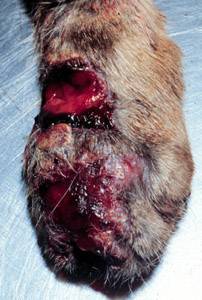
Sporothrix brasiliensis is a fungus that is commonly found in soil. It is an emerging fungal pathogen that is causing disease in humans and cats mainly in Brazil and other countries in South America.

References

  1. 1 2 3 4 5 6 7 8 9 10 11 12 13 14 15 16 17 18 19 20 21 22 Milner, Dan A.; Solomon, Isaac (2020). "Sporotrichosis". In Milner, Danny A. (ed.). Diagnostic Pathology: Infectious Diseases. Elsevier. pp. 316–319. ISBN   978-0-323-61138-1.
  2. 1 2 3 Proia, Laurie (2020). "28. The dimorphic mycoses". In Spec, Andrej; Escota, Gerome V.; Chrisler, Courtney; Davies, Bethany (eds.). Comprehensive Review of Infectious Diseases. Elsevier. pp. 421–422. ISBN   978-0-323-56866-1.
  3. 1 2 3 4 5 James, William D.; Elston, Dirk; Treat, James R.; Rosenbach, Misha A.; Neuhaus, Isaac (2019). "13. Diseases resulting from fungi and yeasts". Andrews' Diseases of the Skin: Clinical Dermatology (13th ed.). Elsevier. pp. 314–315. ISBN   978-0-323-54753-6.
  4. Johnstone, Ronald B. (2017). "25. Mycoses and Algal infections". Weedon's Skin Pathology Essentials (2nd ed.). Elsevier. p. 455. ISBN   978-0-7020-6830-0.
  5. "ICD-11 - ICD-11 for Mortality and Morbidity Statistics". icd.who.int. Retrieved 11 July 2021.
  6. López-Romero, Everardo; et al. (2010). "Sporothrix schenckii complex and sporotrichosis, an emerging health problem". Future Microbiology. 6 (1): 85–102. doi:10.2217/fmb.10.157. PMID   21162638.
  7. Ryan KJ, Ray CG, eds. (2004). Sherris Medical Microbiology (4th ed.). McGraw Hill. pp.  654–6. ISBN   978-0-8385-8529-0.
  8. Barros MB, de Almeida Paes R, Schubach AO (October 2011). "Sporothrix schenckii and Sporotrichosis". Clinical Microbiology Reviews. 24 (4): 633–54. doi:10.1128/CMR.00007-11. PMC   3194828 . PMID   21976602.
  9. 1 2 Procop, Gary W.; et al. (2020). Koneman's Color Atlas and Textbook of Diagnostic Microbiology. Jones & Bartlett Learning. p. 1377. ISBN   9781284322378.
  10. 1 2 3 Birchard, Stephen J.; Sherding, Robert G. (2005). Saunders Manual of Small Animal Practice (3rd ed.). Elsevier Health Sciences. p. 440. ISBN   9781416064299.
  11. 1 2 Sykes, Jane E. (2013). Canine and Feline Infectious Diseases. Elsevier Health Sciences. p. 629. ISBN   9780323241946.
  12. 1 2 Sykes, Jane E. (2022). Greene's Infectious Diseases of the Dog and Cat. Elsevier Health Sciences. p. 1054. ISBN   9780323509336.
  13. Lortholary O, Denning DW, Dupont B (March 1999). "Endemic mycoses: a treatment update". The Journal of Antimicrobial Chemotherapy. 43 (3): 321–31. doi: 10.1093/jac/43.3.321 . PMID   10223586.
  14. Xue S, Gu R, Wu T, Zhang M, Wang X (October 2009). "Oral potassium iodide for the treatment of sporotrichosis". The Cochrane Database of Systematic Reviews. 2009 (4): CD006136. doi:10.1002/14651858.CD006136.pub2. PMC   7388325 . PMID   19821356.
  15. 1 2 Hogan BK, Hospenthal DR (March 2011). "Update on the therapy for sporotrichosis". Patient Care. 22: 49–52. Archived from the original on 2020-05-22. Retrieved 2019-05-05.
  16. Chapman SW, Pappas P, Kauffmann C, Smith EB, Dietze R, Tiraboschi-Foss N, Restrepo A, Bustamante AB, Opper C, Emady-Azar S, Bakshi R (February 2004). "Comparative evaluation of the efficacy and safety of two doses of terbinafine (500 and 1000 mg day(-1)) in the treatment of cutaneous or lymphocutaneous sporotrichosis". Mycoses. 47 (1–2): 62–8. doi:10.1046/j.1439-0507.2003.00953.x. hdl: 2027.42/74074 . PMID   14998402. S2CID   7319396.
  17. Takahashi S, Masahashi T, Maie O (October 1981). "[Local thermotherapy in sporotrichosis]". Der Hautarzt; Zeitschrift für Dermatologie, Venerologie, und Verwandte Gebiete (in German). 32 (10): 525–8. PMID   7298332.
  18. "Sporotrichosis". The Merck Veterinary Manual. Retrieved 2019-06-17.
  19. Carpouron, Julia E.; et al. (2022). "Emerging Animal-Associated Fungal Diseases". Journal of Fungi. 8 (6): 611. doi: 10.3390/jof8060611 . PMC   9225262 . PMID   35736094.