Dermatophytosis

Last updated

Dermatophytosis
Other namesRingworm, tinea
Yeartinfection.JPG
Ringworm on a human leg
Specialty Dermatology, Internal Medicine
Symptoms Red, itchy, scaly, circular skin rash [1]
Causes Fungal infection [2]
Risk factors Using public showers, contact sports, excessive sweating, contact with animals, obesity, poor immune function [3] [4]
Diagnostic method Based on symptoms, microbial culture, microscopic examination [5]
Differential diagnosis Dermatitis, psoriasis, pityriasis rosea, tinea versicolor [6]
PreventionKeep the skin dry, not walking barefoot in public, not sharing personal items [3]
Treatment Antifungal creams (clotrimazole, miconazole) [7]
Frequency20% of the population [8]

Dermatophytosis, also known as ringworm, is a fungal infection of the skin (Dermatomycosis). [2] Typically it results in a red, itchy, scaly, circular rash. [1] Hair loss may occur in the area affected. [1] Symptoms begin four to fourteen days after exposure. [1] Multiple areas can be affected at a given time. [4]

Contents

About 40 types of fungus can cause ringworm. [2] They are typically of the Trichophyton , Microsporum , or Epidermophyton type. [2] Risk factors include using public showers, contact sports such as wrestling, excessive sweating, contact with animals, obesity, and poor immune function. [3] [4] Ringworm can spread from other animals or between people. [3] Diagnosis is often based on the appearance and symptoms. [5] It may be confirmed by either culturing or looking at a skin scraping under a microscope. [5]

Prevention is by keeping the skin dry, not walking barefoot in public, and not sharing personal items. [3] Treatment is typically with antifungal creams such as clotrimazole or miconazole. [7] If the scalp is involved, antifungals by mouth such as fluconazole may be needed. [7]

Globally, up to 20% of the population may be infected by ringworm at any given time. [8] Infections of the groin are more common in males, while infections of the scalp and body occur equally in both sexes. [4] Infections of the scalp are most common in children while infections of the groin are most common in the elderly. [4] Descriptions of ringworm date back to ancient history. [9]

Signs and symptoms

Infections on the body may give rise to typical enlarging raised red rings of ringworm. Infection on the skin of the feet may cause athlete's foot and in the groin, jock itch. Involvement of the nails is termed onychomycosis.

Animals including dogs and cats can also be affected by ringworm, and the disease can be transmitted between animals and humans, making it a zoonotic disease.

Specific signs can be:

Causes

Fungi thrive in moist, warm areas, such as locker rooms, tanning beds, swimming pools, and skin folds; accordingly, those that cause dermatophytosis may be spread by using exercise machines that have not been disinfected after use, or by sharing towels, clothing, footwear, or hairbrushes.

Diagnosis

Dermatophyte infections can be readily diagnosed based on the history, physical examination, and potassium hydroxide (KOH) microscopy. [10]

Classification

Three types of tinea (capitis, corporis, and barbae) Fox Plate LXXXIX.jpg
Three types of tinea (capitis, corporis, and barbae)

A number of different species of fungus are involved in dermatophytosis. Dermatophytes of the genera Trichophyton and Microsporum are the most common causative agents. These fungi attack various parts of the body and lead to the conditions listed below. The Latin names are for the conditions (disease patterns), not the agents that cause them. The disease patterns below identify the type of fungus that causes them only in the cases listed:

Prevention

Advice often given includes:

Vaccination

As of 2016, no approved human vaccine exist against dermatophytosis. For horses, dogs and cats there is available an approved inactivated vaccine called Insol Dermatophyton (Boehringer Ingelheim) which provides time-limited protection against several trichophyton and microsporum fungal strains. [14] With cattle, systemic vaccination has achieved effective control of ringworm. Since 1979 a Russian live vaccine (LFT 130) and later on a Czechoslovakian live vaccine against bovine ringworm has been used. In Scandinavian countries vaccination programmes against ringworm are used as a preventive measure to improve the hide quality. In Russia, fur-bearing animals (silver fox, foxes, polar foxes) and rabbits have also been treated with vaccines. [15]

Treatment

Antifungal treatments include topical agents such as miconazole, terbinafine, clotrimazole, ketoconazole, or tolnaftate applied twice daily until symptoms resolve — usually within one or two weeks. [16] Topical treatments should then be continued for a further 7 days after resolution of visible symptoms to prevent recurrence. [16] [17] The total duration of treatment is therefore generally two weeks, [18] [19] but may be as long as three. [20]

In more severe cases or scalp ringworm, systemic treatment with oral medications (such as itraconazole, terbinafine, and ketoconazole) may be given. [21]

To prevent spreading the infection, lesions should not be touched, and good hygiene maintained with washing of hands and the body. [22]

Misdiagnosis and treatment of ringworm with a topical steroid, a standard treatment of the superficially similar pityriasis rosea, can result in tinea incognito, a condition where ringworm fungus grows without typical features, such as a distinctive raised border.[ citation needed ]

History

Dermatophytosis has been prevalent since before 1906, at which time ringworm was treated with compounds of mercury or sometimes sulfur or iodine. Hairy areas of skin were considered too difficult to treat, so the scalp was treated with X-rays and followed up with antifungal medication. [23] Another treatment from around the same time was application of Araroba powder. [24]

Terminology

The most common term for the infection, "ringworm", is a misnomer, since the condition is caused by fungi of several different species and not by parasitic worms.

Other animals

Ringworm caused by Trichophyton verrucosum is a frequent clinical condition in cattle. Young animals are more frequently affected. The lesions are located on the head, neck, tail, and perineum. [25] The typical lesion is a round, whitish crust. Multiple lesions may coalesce in "map-like" appearance.

Clinical dermatophytosis is also diagnosed in sheep, dogs, cats, and horses. Causative agents, besides Trichophyton verrucosum, areT. mentagrophytes, T. equinum, Microsporum gypseum, M. canis, and M. nanum. [26]

Dermatophytosis may also be present in the holotype of the Cretaceous eutriconodont mammal Spinolestes , suggesting a Mesozoic origin for this disease.

Diagnosis

Ringworm in pets may often be asymptomatic, resulting in a carrier condition which infects other pets. In some cases, the disease only appears when the animal develops an immunodeficiency condition. Circular bare patches on the skin suggest the diagnosis, but no lesion is truly specific to the fungus. Similar patches may result from allergies, sarcoptic mange, and other conditions. Three species of fungi cause 95% of dermatophytosis in pets:[ citation needed ] these are Microsporum canis , Microsporum gypseum , and Trichophyton mentagrophytes .

Veterinarians have several tests to identify ringworm infection and identify the fungal species that cause it:

Woods test: This is an ultraviolet light with a magnifying lens. Only 50% of M. canis will show up as an apple-green fluorescence on hair shafts, under the UV light. The other fungi do not show. The fluorescent material is not the fungus itself (which does not fluoresce), but rather an excretory product of the fungus which sticks to hairs. Infected skin does not fluoresce.

Microscopic test: The veterinarian takes hairs from around the infected area and places them in a staining solution to view under the microscope. Fungal spores may be viewed directly on hair shafts. This technique identifies a fungal infection in about 40%–70% of the infections, but cannot identify the species of dermatophyte.

Culture test: This is the most effective, but also the most time-consuming, way to determine if ringworm is on a pet. In this test, the veterinarian collects hairs from the pet, or else collects fungal spores from the pet's hair with a toothbrush, or other instrument, and inoculates fungal media for culture. These cultures can be brushed with transparent tape and then read by the veterinarian using a microscope, or can be sent to a pathological lab. The three common types of fungi which commonly cause pet ringworm can be identified by their characteristic spores. These are different-appearing macroconidia in the two common species of Microspora, and typical microconidia in Trichophyton infections. [26]

Identifying the species of fungi involved in pet infections can be helpful in controlling the source of infection. M. canis, despite its name, occurs more commonly in domestic cats, and 98% of cat infections are with this organism.[ citation needed ] It can also infect dogs and humans, however. T. mentagrophytes has a major reservoir in rodents, but can also infect pet rabbits, dogs, and horses. M. gypseum is a soil organism and is often contracted from gardens and other such places. Besides humans, it may infect rodents, dogs, cats, horses, cattle, and swine. [27]

Treatment

Pet animals

Treatment requires both systemic oral treatment with most of the same drugs used in humans—terbinafine, fluconazole, or itraconazole—as well as a topical "dip" therapy. [28]

Because of the usually longer hair shafts in pets compared to those of humans, the area of infection and possibly all of the longer hair of the pet must be clipped to decrease the load of fungal spores clinging to the pet's hair shafts. However, close shaving is usually not done because nicking the skin facilitates further skin infection.

Twice-weekly bathing of the pet with diluted lime sulfur dip solution is effective in eradicating fungal spores. This must continue for 3 to 8 weeks. [29]

Washing of household hard surfaces with 1:10 household sodium hypochlorite bleach solution is effective in killing spores, but it is too irritating to be used directly on hair and skin.

Pet hair must be rigorously removed from all household surfaces, and then the vacuum cleaner bag, and perhaps even the vacuum cleaner itself, discarded when this has been done repeatedly. Removal of all hair is important, since spores may survive 12 months or even as long as two years on hair clinging to surfaces. [30]

Cattle

In bovines, an infestation is difficult to cure, as systemic treatment is uneconomical. Local treatment with iodine compounds is time-consuming, as it needs scraping of crusty lesions. Moreover, it must be carefully conducted using gloves, lest the worker become infested.

Epidemiology

Worldwide, superficial fungal infections caused by dermatophytes are estimated to infect around 20-25% of the population and it is thought that dermatophytes infect 10-15% of the population during their lifetime. [31] [32] The highest incidence of superficial mycoses result from dermatophytoses which are most prevalent in tropical regions. [31] [33] Onychomycosis, a common infection caused by dermatophytes, is found with varying prevalence rates in many countries. [34] Tinea pedis + onychomycosis, Tinea corporis, Tinea capitis are the most common dermatophytosis found in humans across the world. [34] Tinea capitis has a greater prevalence in children. [31] The increasing prevalence of dermatophytes resulting in Tinea capitis has been causing epidemics throughout Europe and America. [34] In pets, cats are the most affected by dermatophytosis. [35] Pets are susceptible to dermatophytoses caused by Microsporum canis, Microsporum gypseum, and Trichophyton. [35] For dermatophytosis in animals, risk factors depend on age, species, breed, underlying conditions, stress, grooming, and injuries. [35]

Numerous studies have found Tinea capitis to be the most prevalent dermatophyte to infect children across the continent of Africa. [32] Dermatophytosis has been found to be most prevalent in children ages 4 to 11, infecting more males than females. [32] Low socioeconomic status was found to be a risk factor for Tinea capitis. [32] Throughout Africa, dermatophytoses are common in hot- humid climates and with areas of overpopulation. [32]

Chronicity is a common outcome for dermatophytosis in India. [33] The prevalence of dermatophytosis in India is between 36.6 and 78.4% depending on the area, clinical subtype, and dermatophyte isolate. [33] Individuals ages 21–40 years are most commonly affected. [33]

A 2002 study looking at 445 samples of dermatophytes in patients in Goiânia, Brazil found the most prevalent type to be Trichophyton rubrum (49.4%), followed by Trichophyton mentagrophytes (30.8%), and Microsporum canis (12.6%). [36]

A 2013 study looking at 5,175 samples of Tinea in patients in Tehran, Iran found the most prevalent type to be Tinea pedis (43.4%), followed by Tinea unguium. (21.3%), and Tinea cruris (20.7%). [37]

See also

Related Research Articles

<span class="mw-page-title-main">Athlete's foot</span> Skin infection caused by fungus

Athlete's foot, known medically as tinea pedis, is a common skin infection of the feet caused by a fungus. Signs and symptoms often include itching, scaling, cracking and redness. In rare cases the skin may blister. Athlete's foot fungus may infect any part of the foot, but most often grows between the toes. The next most common area is the bottom of the foot. The same fungus may also affect the nails or the hands. It is a member of the group of diseases known as tinea.

<span class="mw-page-title-main">Griseofulvin</span> Antifungal medication used for dermatophytoses

Griseofulvin is an antifungal medication used to treat a number of types of dermatophytoses (ringworm). This includes fungal infections of the nails and scalp, as well as the skin when antifungal creams have not worked. It is taken by mouth.

Dermatophyte is a common label for a group of fungus of Arthrodermataceae that commonly causes skin disease in animals and humans. Traditionally, these anamorphic mold genera are: Microsporum, Epidermophyton and Trichophyton. There are about 40 species in these three genera. Species capable of reproducing sexually belong in the teleomorphic genus Arthroderma, of the Ascomycota. As of 2019 a total of nine genera are identified and new phylogenetic taxonomy has been proposed.

<span class="mw-page-title-main">Tinea corporis</span> Medical condition

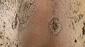
Tinea corporis is a fungal infection of the body, similar to other forms of tinea. Specifically, it is a type of dermatophytosis that appears on the arms and legs, especially on glabrous skin; however, it may occur on any superficial part of the body.

<span class="mw-page-title-main">Tinea capitis</span> Cutaneous fungal infection of the scalp

Tinea capitis is a cutaneous fungal infection (dermatophytosis) of the scalp. The disease is primarily caused by dermatophytes in the genera Trichophyton and Microsporum that invade the hair shaft. The clinical presentation is typically single or multiple patches of hair loss, sometimes with a 'black dot' pattern, that may be accompanied by inflammation, scaling, pustules, and itching. Uncommon in adults, tinea capitis is predominantly seen in pre-pubertal children, more often boys than girls.

<span class="mw-page-title-main">Kerion</span> Medical condition

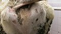
Kerion or kerion celsi is an acute inflammatory process which is the result of the host's response to a fungal ringworm infection of the hair follicles of the scalp that can be accompanied by secondary bacterial infection(s). It usually appears as raised, spongy lesions, and typically occurs in children. This honeycomb is a painful inflammatory reaction with deep suppurative lesions on the scalp. Follicles may be seen discharging pus. There may be sinus formation and rarely mycetoma-like grains are produced. It is usually caused by dermatophytes such as Trichophyton verrucosum, T. mentagrophytes, and Microsporum canis. Treatment with oral griseofulvin common.

<span class="mw-page-title-main">Tinea manuum</span> Medical condition

Tinea manuum is a fungal infection of the hand, mostly a type of dermatophytosis, often part of two feet-one hand syndrome. There is diffuse scaling on the palms or back of usually one hand and the palmer creases appear more prominent. When both hands are affected, the rash looks different on each hand, with palmer creases appearing whitish if the infection has been present for a long time. It can be itchy and look slightly raised. Nails may also be affected.

<span class="mw-page-title-main">KOH test</span> Test to differentiate between skin fungi

The KOH Test for Candida albicans, also known as a potassium hydroxide preparation or KOH prep, is a quick, inexpensive fungal test to differentiate dermatophytes and Candida albicans symptoms from other skin disorders like psoriasis and eczema.

<i>Trichophyton</i> Genus of Fungi

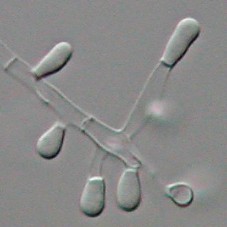
Trichophyton is a genus of fungi, which includes the parasitic varieties that cause tinea, including athlete's foot, ringworm, jock itch, and similar infections of the nail, beard, skin and scalp. Trichophyton fungi are molds characterized by the development of both smooth-walled macro- and microconidia. Macroconidia are mostly borne laterally directly on the hyphae or on short pedicels, and are thin- or thick-walled, clavate to fusiform, and range from 4 to 8 by 8 to 50 μm in size. Macroconidia are few or absent in many species. Microconidia are spherical, pyriform to clavate or of irregular shape, and range from 2 to 3 by 2 to 4 μm in size.

<i>Microsporum audouinii</i> Species of fungus

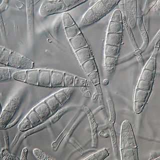
Microsporum audouinii is an anthropophilic fungus in the genus Microsporum. It is a type of dermatophyte that colonizes keratinized tissues causing infection. The fungus is characterized by its spindle-shaped macroconidia, clavate microconidia as well as its pitted or spiny external walls.

<i>Trichophyton tonsurans</i> Species of fungus

Trichophyton tonsurans is a fungus in the family Arthrodermataceae that causes ringworm infection of the scalp. It was first recognized by David Gruby in 1844. Isolates are characterized as the "–" or negative mating type of the Arthroderma vanbreuseghemii complex. This species is thought to be conspecific with T. equinum, although the latter represents the "+" mating strain of the same biological species Despite their biological conspecificity, clones of the two mating types appear to have undergone evolutionary divergence with isolates of the T. tonsurans-type consistently associated with Tinea capitis whereas the T. equinum-type, as its name implies, is associated with horses as a regular host. Phylogenetic relationships were established in isolates from Northern Brazil, through fingerprinting polymorphic RAPD and M13 markers. There seems to be lower genomic variability in the T. tonsurans species due to allopatric divergence. Any phenotypic density is likely due to environmental factors, not genetic characteristics of the fungus.

<span class="mw-page-title-main">Fungal folliculitis</span> Inflammation of hair follicles due to fungal infection

Majocchi's granuloma is a skin condition characterized by deep, pustular plaques, and is a form of tinea corporis. It is a localized form of fungal folliculitis. Lesions often have a pink and scaly central component with pustules or folliculocentric papules at the periphery. The name comes from Domenico Majocchi, who discovered the disorder in 1883. Majocchi was a professor of dermatology at the University of Parma and later the University of Bologna. The most common dermatophyte is called Trichophyton rubrum.

<i>Microsporum gypseum</i> Species of fungus

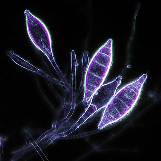
Microsporum gypseum is a soil-associated dermatophyte that occasionally is known to colonise and infect the upper dead layers of the skin of mammals. The name refers to an asexual "form-taxon" that has been associated with four related biological species of fungi: the pathogenic taxa Arthroderma incurvatum, A. gypsea, A. fulva and the non-pathogenic saprotroph A. corniculata. More recent studies have restricted M. gypseum to two teleomorphic species A. gypseum and A. incurvatum. The conidial states of A. fulva and A. corniculata have been assigned to M. fulvum and M. boullardii. Because the anamorphic states of these fungi are so similar, they can be identified reliably only by mating. Two mating strains have been discovered, "+" and "–". The classification of this species has been based on the characteristically rough-walled, blunt, club-shaped, multicelled macroconidia. Synonyms include Achorion gypseum, Microsporum flavescens, M. scorteum, and M. xanthodes. There has been past nomenclatural confusion in the usage of the generic names Microsporum and Microsporon.

<i>Microsporum canis</i> Species of fungus

Microsporum canis is a pathogenic, asexual fungus in the phylum Ascomycota that infects the upper, dead layers of skin on domesticated cats, and occasionally dogs and humans. The species has a worldwide distribution.

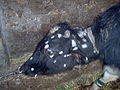
Microsporum nanum is a pathogenic fungus in the family Arthrodermataceae. It is a type of dermatophyte that causes infection in dead keratinized tissues such as skin, hair, and nails. Microsporum nanum is found worldwide and is both zoophilic and geophilic. Animals such as pigs and sheep are the natural hosts for the fungus; however, infection of humans is also possible. Majority of the human cases reported are associated with pig farming. The fungus can invade the skin of the host; if it is scratched off by the infected animal, the fungus is still capable of reproducing in soil.

<i>Microsporum gallinae</i> Species of fungus

Microsporum gallinae is a fungus of the genus Microsporum that causes dermatophytosis, commonly known as ringworm. Chickens represent the host population of Microsporum gallinae but its opportunistic nature allows it to enter other populations of fowl, mice, squirrels, cats, dogs and monkeys. Human cases of M. gallinae are rare, and usually mild, non-life-threatening superficial infections.

<span class="mw-page-title-main">Favus</span> Medical condition

Favus or tinea favosa is the severe form of tinea capitis, a skin infectious disease caused by the dermatophyte fungus Trichophyton schoenleinii. Typically the species affects the scalp, but occasionally occurs as onychomycosis, tinea barbae, or tinea corporis.

<i>Epidermophyton floccosum</i> Species of fungus

Epidermophyton floccosum is a filamentous fungus that causes skin and nail infections in humans. This anthropophilic dermatophyte can lead to diseases such as tinea pedis, tinea cruris, tinea corporis and onychomycosis. Diagnostic approaches of the fungal infection include physical examination, culture testing, and molecular detection. Topical antifungal treatment, such as the use of terbinafine, itraconazole, voriconazole, and ketoconazole, is often effective.

Microsporum fulvum is a wildly-distributed dermatophyte species in the Fungi Kingdom. It is known to be a close relative to other dermatophytes such as Trichophyton andEpidermophyton. The fungus is common within soil environments and grows well on keratinized material, such as hair, nails and dead skin. It is recognized as an opportunistic fungal pathogen capable of causing cutaneous mycoses in humans and animals. Originally, the fungus was thought to be Microsporum gypseum until enhanced genetic examination separated the two as distinct species in 1963.

References

  1. 1 2 3 4 "Symptoms of Ringworm Infections". CDC. December 6, 2015. Archived from the original on 20 January 2016. Retrieved 5 September 2016.
  2. 1 2 3 4 "Definition of Ringworm". CDC. December 6, 2015. Archived from the original on 5 September 2016. Retrieved 5 September 2016.
  3. 1 2 3 4 5 "Ringworm Risk & Prevention". CDC. December 6, 2015. Archived from the original on 7 September 2016. Retrieved 5 September 2016.
  4. 1 2 3 4 5 Domino FJ, Baldor RA, Golding J (2013). The 5-Minute Clinical Consult 2014. Lippincott Williams & Wilkins. p. 1226. ISBN   9781451188509. Archived from the original on 2016-09-15.
  5. 1 2 3 "Diagnosis of Ringworm". CDC. December 6, 2015. Archived from the original on 8 August 2016. Retrieved 5 September 2016.
  6. Teitelbaum JE (2007). In a Page: Pediatrics. Lippincott Williams & Wilkins. p. 274. ISBN   9780781770453. Archived from the original on 2017-04-26.
  7. 1 2 3 "Treatment for Ringworm". CDC. December 6, 2015. Archived from the original on 3 September 2016. Retrieved 5 September 2016.
  8. 1 2 Mahmoud A. Ghannoum, John R. Perfect (24 November 2009). Antifungal Therapy. CRC Press. p. 258. ISBN   978-0-8493-8786-9. Archived from the original on 8 September 2017.
  9. Bolognia JL, Jorizzo JL, Schaffer JV (2012). Dermatology (3 ed.). Elsevier Health Sciences. p. 1255. ISBN   978-0702051821. Archived from the original on 2016-09-15.
  10. Barry L. Hainer, M.D., Medical University of South Carolina, Charleston, South Carolina (2003). "Dermatophyte Infections". Am Fam Physician. 67 (1): 101–109. PMID   12537173.{{cite journal}}: CS1 maint: multiple names: authors list (link)
  11. Klemm L (2 April 2008). "Keeping footloose on trips". The Herald News. Archived from the original on 18 February 2009.
  12. Fort Dodge Animal Health: Milestones from Wyeth.com. Retrieved April 28, 2008.
  13. "Ringworm In Your Dog, Cat And Other Pets". Vetspace. Retrieved 14 November 2020.
  14. "Insol Dermatophyton 5x2 ml". GROVET - The veterinary warehouse. Archived from the original on 2016-08-17. Retrieved 2016-02-01.
  15. F. Rochette, M. Engelen, H. Vanden Bossche (2003), "Antifungal agents of use in animal health - practical applications", Journal of Veterinary Pharmacology and Therapeutics , 26 (1): 31–53, doi:10.1046/j.1365-2885.2003.00457.x, PMID   12603775
  16. 1 2 Kyle AA, Dahl MV (2004). "Topical therapy for fungal infections". Am J Clin Dermatol. 5 (6): 443–51. doi:10.2165/00128071-200405060-00009. PMID   15663341. S2CID   37500893.
  17. McClellan KJ, Wiseman LR, Markham A (July 1999). "Terbinafine. An update of its use in superficial mycoses". Drugs. 58 (1): 179–202. doi:10.2165/00003495-199958010-00018. PMID   10439936. S2CID   195691703.
  18. Tinea~treatment at eMedicine
  19. Tinea Corporis~treatment at eMedicine
  20. "Antifungal agents for common paediatric infections". Can J Infect Dis Med Microbiol. 19 (1): 15–8. January 2008. doi: 10.1155/2008/186345 . PMC   2610275 . PMID   19145261.
  21. Gupta AK, Cooper EA (2008). "Update in antifungal therapy of dermatophytosis". Mycopathologia. 166 (5–6): 353–67. doi:10.1007/s11046-008-9109-0. PMID   18478357. S2CID   24116721.
  22. "Ringworm on Body Treatment" at eMedicineHealth
  23. Sequeira, J.H. (1906). "The Varieties of Ringworm and Their Treatment" (PDF). British Medical Journal. 2 (2378): 193–196. doi:10.1136/bmj.2.2378.193. PMC   2381801 . PMID   20762800. Archived (PDF) from the original on 2009-11-22.
  24. Mrs. M. Grieve. A Modern Herbal. Archived from the original on 2015-03-25.
  25. Scott DW (2007). Colour Atlas of Animal Dermatology. Blackwell. ISBN   978-0-8138-0516-0.
  26. 1 2 David W. Scott, Colour Atlas of Animal Dermatology, Blackwell Publishing Professional 2121 State Avenue, Ames, Iowa 50014, USA; ISBN 978-0-8138-0516-0/2007.
  27. "General ringworm information". Ringworm.com.au. Archived from the original on 2010-12-21. Retrieved 2011-01-10.
  28. "Facts About Ringworm". Archived from the original on 2011-10-06. Retrieved 2011-10-03. Detailed veterinary discussion of animal treatment
  29. "Veterinary treatment site page". Marvistavet.com. Archived from the original on 2013-01-04. Retrieved 2011-01-10.
  30. "Persistence of spores". Ringworm.com.au. Archived from the original on 2010-12-21. Retrieved 2011-01-10.
  31. 1 2 3 Pires, C. A. A., Cruz, N. F. S. da, Lobato, A. M., Sousa, P. O. de, Carneiro, F. R. O., & Mendes, A. M. D. (2014). Clinical, epidemiological, and therapeutic profile of dermatophytosis. Anais Brasileiros de Dermatología, 89(2), 259–264.
  32. 1 2 3 4 5 Oumar Coulibaly, Coralie L'Ollivier, Renaud Piarroux, Stéphane Ranque, Epidemiology of human dermatophytoses in Africa, Medical Mycology, Volume 56, Issue 2, February 2018, Pages 145–161.
  33. 1 2 3 4 Rajagopalan, M., Inamadar, A., Mittal, A., Miskeen, A. K., Srinivas, C. R., Sardana, K., Godse, K., Patel, K., Rengasamy, M., Rudramurthy, S., & Dogra, S. (2018). Expert Consensus on The Management of Dermatophytosis in India (ECTODERM India). BMC dermatology, 18(1), 6.
  34. 1 2 3 Hayette, M.-P., & Sacheli, R. (2015). Dermatophytosis, Trends in Epidemiology and Diagnostic Approach. Current Fungal Infection Reports, 9(3), 164–179.
  35. 1 2 3 Gordon, E., Idle, A., & DeTar, L. (2020). Descriptive epidemiology of companion animal dermatophytosis in a Canadian Pacific Northwest animal shelter system. The Canadian veterinary journal = La revue veterinaire canadienne, 61(7), 763–770.
  36. Costa, M., Passos, X. S., Hasimoto e Souza, L. K., Miranda, A. T. B., Lemos, J. de A., Oliveira, J., & Silva, M. do R. R. (2002). Epidemiology and etiology of dermatophytosis in Goiânia, GO, Brazil. Revista da Sociedade Brasileira de Medicina Tropical, 35(1), 19–.
  37. Rezaei-Matehkolaei, A., Makimura, K., de Hoog, S., Shidfar, M. R., Zaini, F., Eshraghian, M., Naghan, P. A., & Mirhendi, H. (2013). Molecular epidemiology of dermatophytosis in Tehran, Iran, a clinical and microbial survey. Medical Mycology (Oxford), 51(2), 203–207.

Further reading