Cryptococcus neoformans

Last updated

Contents

Cryptococcus neoformans
Cryptococcus neoformans using a light India ink staining preparation PHIL 3771 lores.jpg
Yeast state of Cryptococcus neoformans
Scientific classification OOjs UI icon edit-ltr.svg
Domain: Eukaryota
Kingdom: Fungi
Division: Basidiomycota
Class: Tremellomycetes
Order: Tremellales
Family: Cryptococcaceae
Genus: Cryptococcus
Species:
C. neoformans
Binomial name
Cryptococcus neoformans
(San Felice) Vuill. (1901)
Synonyms

Saccharomyces neoformansSan Felice (1895)
Filobasidiella neoformansKwon-Chung (1975)

Cryptococcus neoformans is an encapsulated yeast [1] belonging to the class Tremellomycetes and an obligate aerobe [2] that can live in both plants and animals. Its teleomorph is a filamentous fungus, formerly referred to Filobasidiella neoformans. In its yeast state, it is often found in bird excrement. Cryptococcus neoformans can cause disease in apparently immunocompetent, as well as immunocompromised, hosts. [1] [3]

Classification

Cryptococcus neoformans has undergone numerous nomenclature revisions since its first description in 1895. It formerly contained two varieties: C. neoformans var. neoformans and C. neoformans var. grubii. A third variety, C. neoformans var. gattii, was later defined as a distinct species, Cryptococcus gattii . The most recent classification system divides these varieties into seven species. [4] C. neoformans refers to C. neoformans var. grubii. A new species name, Cryptococcus deneoformans, is used for the former C. neoformans var. neoformans. C. gattii is divided into five species.[ citation needed ]

The teleomorph was first described in 1975 by K.J. Kwon-Chung, who obtained cultures of Filobasidiella neoformans by crossing strains of the yeast C. neoformans. She was able to observe basidia similar to those of the genus Filobasidium , hence the name Filobasidiella for the new genus. [5] Following changes to the International Code of Nomenclature for algae, fungi, and plants, the practice of giving different names to teleomorph and anamorph forms of the same fungus was discontinued, meaning that Filobasidiella neoformans became a synonym of the earlier name Cryptococcus neoformans.[ citation needed ]

Characteristics

C. neoformans stained by Gram stain Cryptococcus Gram film.jpg
C. neoformans stained by Gram stain

Cryptococcus neoformans typically grows as a yeast (unicellular) and replicates by budding. It makes hyphae during mating, and eventually creates basidiospores at the end of the hyphae before producing spores. Under host-relevant conditions, including low glucose, serum, 5% carbon dioxide, and low iron, among others, the cells produce a characteristic polysaccharide capsule. [6] The recognition of C. neoformans in Gram-stained smears of purulent exudates may be hampered by the presence of the large gelatinous capsule which apparently prevents definitive staining of the yeast-like cells. In such stained preparations, it may appear either as round cells with Gram-positive granular inclusions impressed upon a pale lavender cytoplasmic background or as Gram-negative lipoid bodies. [7] When grown as a yeast, C. neoformans has a prominent capsule composed mostly of polysaccharides. Under the microscope, the India ink stain is used for easy visualization of the capsule in cerebral spinal fluid. [8] The particles of ink pigment do not enter the capsule that surrounds the spherical yeast cell, resulting in a zone of clearance or "halo" around the cells. This allows for quick and easy identification of C. neoformans. Unusual morphological forms are rarely seen. [9] For identification in tissue, mucicarmine stain provides specific staining of polysaccharide cell wall in C. neoformans. Cryptococcal antigen from cerebrospinal fluid is thought to be the best test for diagnosis of cryptococcal meningitis in terms of sensitivity, though it might be unreliable in HIV-positive patients. [10]

The first genome sequence for a strain of C. neoformans (var. neoformans; now C. deneoformans) was published in 2005. [11]

Studies suggest that colonies of C. neoformans and related fungi growing within the ruins of the Chernobyl Nuclear Power Plant may be able to metabolize ionizing radiation. [12]

Pathology

Infection with C. neoformans is termed cryptococcosis. Most infections with C. neoformans occur in the lungs. [13] However, fungal meningitis and encephalitis, especially as a secondary infection for AIDS patients, are often caused by C. neoformans, making it a particularly dangerous fungus. Infections with this fungus are rare in people with fully functioning immune systems, hence C. neoformans is often referred to as an opportunistic pathogen. [1] It is a facultative intracellular pathogen [14] that can utilize host phagocytes to spread within the body. [15] [16] Cryptococcus neoformans was the first intracellular pathogen for which the non-lytic escape process termed vomocytosis was observed. [17] [18] It has been speculated that this ability to manipulate host cells results from environmental selective pressure by amoebae, a hypothesis first proposed by Arturo Casadevall under the term "accidental virulence". [19]

In human infection, C. neoformans is spread by inhalation of aerosolized basidiospores, and can disseminate to the central nervous system, where it can cause meningoencephalitis. [20] In the lungs, C. neoformans cells are phagocytosed by alveolar macrophages. [21] Macrophages produce oxidative and nitrosative agents, creating a hostile environment, to kill invading pathogens. [22] However, some C. neoformans cells can survive intracellularly in macrophages. [21] Intracellular survival appears to be the basis for latency, disseminated disease, and resistance to eradication by antifungal agents. One mechanism by which C. neoformans survives the hostile intracellular environment of the macrophage involves upregulation of expression of genes involved in responses to oxidative stress. [21]

Traversal of the blood–brain barrier by C. neoformans plays a key role in meningitis pathogenesis. [23] However, precise mechanisms by which it passes the blood-brain barrier are still unknown; one recent study in rats suggested an important role of secreted serine proteases. [24] The metalloprotease Mpr1 has been demonstrated to be critical in blood-brain barrier penetration. [25]

Meiosis (sexual reproduction), another possible survival factor for intracellular C. neoformans

The vast majority of environmental and clinical isolates of C. neoformans are mating type alpha. Filaments of mating type alpha have haploid nuclei ordinarily, but these can undergo a process of diploidization (perhaps by endoduplication or stimulated nuclear fusion) to form diploid cells termed blastospores. The diploid nuclei of blastospores are able to undergo meiosis, including recombination, to form haploid basidiospores that can then be dispersed. [26] This process is referred to as monokaryotic fruiting. Required for this process is a gene designated dmc1, a conserved homologue of genes recA in bacteria, and rad51 in eukaryotes (see articles recA and rad51). Dmc1 mediates homologous chromosome pairing during meiosis and repair of double-strand breaks in DNA. [27] One benefit of meiosis in C. neoformans could be to promote DNA repair in the DNA-damaging environment caused by the oxidative and nitrosative agents produced in macrophages. [26] Thus, C. neoformans can undergo a meiotic process, monokaryotic fruiting, that may promote recombinational repair in the oxidative, DNA-damaging environment of the host macrophage, and this may contribute to its virulence.[ citation needed ]

Serious complications of human infection

Infection starts in lungs, disseminates via blood to meninges and then to other parts of the body. Capsule inhibits phagocytosis. Can cause a systemic infection, including fatal meningitis known as meningoencephalitis in normal, diabetic and immunocompromised hosts. The infection from C. neoformans in the brain can be fatal if untreated. CNS (central nervous system) infection may also be present as a brain abscess known as cryptococcomas, subdural effusion, dementia, isolated cranial nerve lesion, spinal cord lesion, and ischemic stroke. If cryptococcal meningitis occurs, mortality rate is between 10 and 30%. [28] Cryptococcal meningitis causes more than 180000 deaths annually [29] .The estimated one-year mortality of HIV-related people who receive treatment for cryptococcal meningitis is 70% in low-income countries versus 20–30% for high-income countries. [30] The symptoms include headache, fever, neck stiffness, nausea and vomiting, photophobia. The diagnosis methods contain a serum cryptococcal antigen test and lumbar puncture with cerebrospinal fluid (CSF) examination to find out the Cryptococcus neoformans. [31]

Treatment

C. neoformans seen in the lung of a patient with AIDS. Mucicarmine stain is used in this case, staining the inner capsule of the organism red. Cryptococcosis of lung in patient with AIDS. Mucicarmine stain 962 lores.jpg
C. neoformans seen in the lung of a patient with AIDS. Mucicarmine stain is used in this case, staining the inner capsule of the organism red.

Cryptococcosis that does not affect the central nervous system can be treated with fluconazole alone.

Cryptococcal meningitis should be treated for two weeks with intravenous amphotericin B 0.7–1.0 mg/kg/day and oral flucytosine 100 mg/kg/day (or intravenous flucytosine 75 mg/kg/day if the patient is unable to swallow). This should then be followed by oral fluconazole 400–800 mg daily for ten weeks [32] and then 200 mg daily for at least one year and until the patient's CD4 count is above 200 cells/mcl. [33] [34] Flucytosine is a generic, off-patent medicine. However, a market failure exists, with a two-week cost of flucytosine therapy being about $10,000. As a result, flucytosine is currently universally unavailable in low- and middle-income countries. In 1970, flucytosine was available in Africa. [35] The dose of 200 mg/kg/day for flucytosine is not more effective, is associated with more side effects and should not be used. [Needs a reference?]

A single high dose of liposomal amphotericin B with 14 days of flucytosine and fluconazole is recommended by the newest WHO guideline for cryptococcal meningitis. [36] A new study found that brain glucose can trigger amphotericin B (AmB) tolerance of C. neoformans during meningitis which means it needs longer treatment time to kill the fungal cells. The study found that the brain glucose induced AmB tolerance of C. neoformans via glucose repression activator Mig1. Mig1 inhibits the production of ergosterol, the target of AmB, and promotes the production of inositol phosphoryl ceramide (IPC), which competes with AmB for ergosterol to limit AmB efficacy in mouse brain and human CSF. Strikingly, Results of this study indicated that IPC synthase inhibitor aureobasidin A (AbA) can enhance the anti-cryptococcal activity of AmB. AbA+AmB AmB had an even better therapeutic effect in a mouse model of cryptococcal meningitis than AmB+flucytosine which may bring new hope for the treatment of Cryptococcal meningitis. [37]

In Africa, oral fluconazole at a rate of 200 mg daily is often used. However, this does not result in cure, because it merely suppresses the fungus and does not kill it; viable fungus can continue to be grown from cerebrospinal fluid of patients not having taken fluconazole for many months. An increased dose of 400 mg daily does not improve outcomes, [38] but prospective studies from Uganda and Malawi reported that higher doses of 1200 mg per day have more fungicidal activity. [39] The outcomes with fluconazole monotherapy have 30% worse survival than amphotericin-based therapies, in a recent systematic review. [40]

Related Research Articles

<span class="mw-page-title-main">Basidiomycota</span> Division of fungi

Basidiomycota is one of two large divisions that, together with the Ascomycota, constitute the subkingdom Dikarya within the kingdom Fungi. Members are known as basidiomycetes. More specifically, Basidiomycota includes these groups: agarics, puffballs, stinkhorns, bracket fungi, other polypores, jelly fungi, boletes, chanterelles, earth stars, smuts, bunts, rusts, mirror yeasts, and Cryptococcus, the human pathogenic yeast.

<i>Cryptococcus</i> Genus of fungi

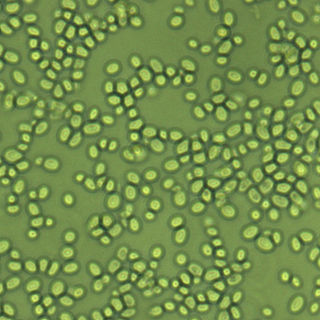
Cryptococcus is a genus of fungi in the family Cryptococcaceae that includes both yeasts and filamentous species. The filamentous, sexual forms or teleomorphs were formerly classified in the genus Filobasidiella, while Cryptococcus was reserved for the yeasts. Most yeast species formerly referred to Cryptococcus have now been placed in different genera. The name Cryptococcus comes from the Greek for "hidden sphere". Some Cryptococcus species cause a disease called cryptococcosis.

<span class="mw-page-title-main">Amphotericin B</span> Antifungal and antiparasitaric chemical compound

Amphotericin B is an antifungal medication used for serious fungal infections and leishmaniasis. The fungal infections it is used to treat include mucormycosis, aspergillosis, blastomycosis, candidiasis, coccidioidomycosis, and cryptococcosis. For certain infections it is given with flucytosine. It is typically given intravenously.

<span class="mw-page-title-main">Cryptococcosis</span> Potentially fatal fungal disease

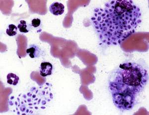
Cryptococcosis is a potentially fatal fungal infection of mainly the lungs, presenting as a pneumonia, and brain, where it appears as a meningitis. Cough, difficulty breathing, chest pain and fever are seen when the lungs are infected. When the brain is infected, symptoms include headache, fever, neck pain, nausea and vomiting, light sensitivity and confusion or changes in behavior. It can also affect other parts of the body including skin, where it may appear as several fluid-filled nodules with dead tissue.

<span class="mw-page-title-main">Fluconazole</span> Antifungal medication

Fluconazole is an antifungal medication used for a number of fungal infections. This includes candidiasis, blastomycosis, coccidioidomycosis, cryptococcosis, histoplasmosis, dermatophytosis, and tinea versicolor. It is also used to prevent candidiasis in those who are at high risk such as following organ transplantation, low birth weight babies, and those with low blood neutrophil counts. It is given either by mouth or by injection into a vein.

<span class="mw-page-title-main">Bacterial capsule</span> Polysaccharide layer that lies outside the cell envelope in many bacteria

The bacterial capsule is a large structure common to many bacteria. It is a polysaccharide layer that lies outside the cell envelope, and is thus deemed part of the outer envelope of a bacterial cell. It is a well-organized layer, not easily washed off, and it can be the cause of various diseases.

Intracellular parasites are microparasites that are capable of growing and reproducing inside the cells of a host. They are also called intracellular pathogens.

<span class="mw-page-title-main">Sporotrichosis</span> Medical condition

Sporotrichosis, also known as rose handler's disease, is a fungal infection that may be localised to skin, lungs, bone and joint, or become systemic. It presents with firm painless nodules that later ulcerate. Following initial exposure to Sporothrix schenckii, the disease typically progresses over a period of a week to several months. Serious complications may develop in people who have a weakened immune system.

<span class="mw-page-title-main">Flucytosine</span> Chemical compound

Flucytosine, also known as 5-fluorocytosine (5-FC), is an antifungal medication. It is specifically used, together with amphotericin B, for serious Candida infections and cryptococcosis. It may be used by itself or with other antifungals for chromomycosis. Flucytosine is used by mouth and by injection into a vein.

Immune reconstitution inflammatory syndrome (IRIS) is a condition seen in some cases of HIV/AIDS or immunosuppression, in which the immune system begins to recover, but then responds to a previously acquired opportunistic infection with an overwhelming inflammatory response that paradoxically makes the symptoms of infection worse.

<i>Nakaseomyces glabratus</i> Species of fungus

Nakaseomyces glabratus is a species of haploid yeast of the genus Nakaseomyces, previously known as Candida glabrata. Despite the fact that no sexual life cycle has been documented for this species, N. glabratus strains of both mating types are commonly found. N. glabrata is generally a commensal of human mucosal tissues, but in today's era of wider human immunodeficiency from various causes, N. glabratus is often the second or third most common cause of candidiasis as an opportunistic pathogen. Infections caused by N. glabratus can affect the urogenital tract or even cause systemic infections by entrance of the fungal cells in the bloodstream (Candidemia), especially prevalent in immunocompromised patients.

<span class="mw-page-title-main">Phagolysosome</span> Cytoplasmic body

In biology, a phagolysosome, or endolysosome, is a cytoplasmic body formed by the fusion of a phagosome with a lysosome in a process that occurs during phagocytosis. Formation of phagolysosomes is essential for the intracellular destruction of microorganisms and pathogens. It takes place when the phagosome's and lysosome's membranes 'collide', at which point the lysosomal contents—including hydrolytic enzymes—are discharged into the phagosome in an explosive manner and digest the particles that the phagosome had ingested. Some products of the digestion are useful materials and are moved into the cytoplasm; others are exported by exocytosis.

<i>Cryptococcus gattii</i> Species of fungus

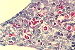
Cryptococcus gattii, formerly known as Cryptococcus neoformans var. gattii, is an encapsulated yeast found primarily in tropical and subtropical climates. Its teleomorph is Filobasidiella bacillispora, a filamentous fungus belonging to the class Tremellomycetes.

Pathogenic fungi are fungi that cause disease in humans or other organisms. Although fungi are eukaryotic, many pathogenic fungi are microorganisms. Approximately 300 fungi are known to be pathogenic to humans; their study is called "medical mycology". Fungal infections are estimated to kill more people than either tuberculosis or malaria—about two million people per year.

<span class="mw-page-title-main">OSU-03012</span> Chemical compound

OSU-03012 (AR-12) is a celecoxib derivative with anticancer and anti-microbial activity. Unlike celecoxib, OSU-03012 does not inhibit COX, but inhibits several other important enzymes instead which may be useful in the treatment of some forms of cancer, When combined with PDE5 inhibitors such as sildenafil or tadalafil, OSU-03012 was found to show enhanced anti-tumour effects in cell culture.

Vomocytosis is the cellular process by phagocytes expel live organisms that they have engulfed without destroying the organism. Vomocytosis is one of many methods used by cells to expel internal materials into their external environment, yet it is distinct in that both the engulfed organism and host cell remain undamaged by expulsion. As engulfed organisms are released without being destroyed, vomocytosis has been hypothesized to be utilized by pathogens as an escape mechanism from the immune system. The exact mechanisms, as well as the repertoire of cells that utilize this mechanism, are currently unknown, yet interest in this unique cellular process is driving continued research with the hopes of elucidating these unknowns.

<span class="mw-page-title-main">Arturo Casadevall</span> Cuban-American scientist

Arturo Casadevall is a Bloomberg Distinguished Professor of Molecular Microbiology & Immunology and Infectious Diseases at the Johns Hopkins Bloomberg School of Public Health and Johns Hopkins School of Medicine, and the Alfred and Jill Sommer Professor and Chair of the W. Harry Feinstone Department of Molecular Microbiology and Immunology at the Johns Hopkins Bloomberg School of Public Health. He is an internationally recognized expert in infectious disease research, with a focus on fungal and bacterial pathogenesis and basic immunology of antibody structure-function. He was elected a member of the National Academy of Sciences in 2022.

<span class="mw-page-title-main">Joseph Heitman</span>

Joseph Heitman is an American physician-scientist focused on research in genetics, microbiology, and infectious diseases. He is the James B. Duke Professor and Chair of the Department of Molecular Genetics and Microbiology at Duke University School of Medicine.

Naganishia albida is a species of fungus in the family Filobasidiaceae. It is currently only known from its yeast state. The species was originally isolated from the air in Japan, and has subsequently been isolated from dry moss in Portugal, grasshoppers in Portugal, and tubercular lungs.

<span class="mw-page-title-main">Chronic meningitis</span> Inflammation of the membranes surrounding the brain and spinal cord lasting longer than 4 weeks

Chronic meningitis is a long-lasting inflammation of the membranes lining the brain and spinal cord. By definition, the duration of signs, symptoms and inflammation in chronic meningitis last longer than 4 weeks. Infectious causes are a leading cause and the infectious organisms responsible for chronic meningitis are different than the organisms that cause acute infectious meningitis. Tuberculosis and the fungi cryptococcus are leading causes worldwide. Chronic meningitis due to infectious causes are more common in those who are immunosuppressed, including those with HIV infection or in children who are malnourished. Chronic meningitis sometimes has a more indolent course than acute meningitis with symptoms developing more insidiously and slowly. Also, some of the infectious agents that cause chronic infectious meningitis such as mycobacterium tuberculosis, many fungal species and viruses are difficult to isolate from the cerebrospinal fluid making diagnosis challenging. No cause is identified during initial evaluation in one third of cases. Magnetic resonance imaging (MRI) of the brain is more sensitive than computed tomography and may show radiological signs that suggest chronic meningitis, however no radiological signs are considered pathognomonic or characteristic. MRI is also normal in many cases further limiting its diagnostic utility.

References

  1. 1 2 3 Buchanan, Kent; Murphy, J. W. (1998). "What makes Cryptococcus neoformans a pathogen?". Emerging Infectious Diseases. 4 (1): 71–83. doi:10.3201/eid0401.980109. PMC   2627665 . PMID   9452400.
  2. Ingavale, Susham S.; Chang, Yun C.; Lee, Hyeseung; McClelland, Carol M.; Leong, Madeline L.; Kwon-Chung, Kyung J. (2008-09-01). "Importance of mitochondria in survival of Cryptococcus neoformans under low oxygen conditions and tolerance to Cobalt Chloride". PLOS Pathogens. 4 (9): e1000155. doi: 10.1371/journal.ppat.1000155 . ISSN   1553-7366. PMC   2528940 . PMID   18802457.
  3. Bahn, Yong-Sun; Sun, Sheng; Heitman, Joseph; Lin, Xiaorong (2020-09-01). "Microbe Profile: Cryptococcus neoformans species complex". Microbiology. 166 (9): 797–799. doi: 10.1099/mic.0.000973 . ISSN   1350-0872. PMC   7717486 . PMID   32956032.
  4. Hagen, Ferry; Khayhan, Kantarawee; Theelen, Bart; Kolecka, Anna; Polacheck, Itzhack; Sionov, Edward; Falk, Rama; Parnmen, Sittiporn; Lumbsch, H. Thorsten (2015-05-01). "Recognition of seven species in the Cryptococcus gattii/Cryptococcus neoformans species complex". Fungal Genetics and Biology. 78: 16–48. doi: 10.1016/j.fgb.2015.02.009 . ISSN   1096-0937. PMID   25721988.
  5. Kwon-Chung KJ. (1975). "A new genus, Filobasidiella, the perfect state of Cryptococcus neoformans". Mycologia. 67 (6): 1197–1200. doi:10.2307/3758842. JSTOR   3758842. PMID   765816.
  6. Heitman, Joseph; Kozel, Thomas R.; Kwon-Chung, Kyung J.; Perfect, John R.; Casadevall, Arturo, eds. (2011). Cryptococcus: From Human Pathogen to Model Yeast. Washington, DC: ASM Press. doi:10.1128/9781555816858. ISBN   9781683671220.
  7. Bottone, E J (1980). "Cryptococcus neoformans: pitfalls in diagnosis through evaluation of gram-stained smears of purulent exudates". Journal of Clinical Microbiology. 12 (6): 790–1. doi:10.1128/jcm.12.6.790-791.1980. PMC   273699 . PMID   6171578.
  8. Zerpa, R; Huicho, L; Guillén, A (September 1996). "Modified India ink preparation for Cryptococcus neoformans in cerebrospinal fluid specimens". Journal of Clinical Microbiology. 34 (9): 2290–1. doi:10.1128/JCM.34.9.2290-2291.1996. PMC   229234 . PMID   8862601.
  9. Shashikala; Kanungo, R; Srinivasan, S; Mathew, R; Kannan, M (Jul–Sep 2004). "Unusual morphological forms of Cryptococcus neoformans in cerebrospinal fluid". Indian Journal of Medical Microbiology. 22 (3): 188–90. doi: 10.1016/S0255-0857(21)02835-8 . PMID   17642731.
  10. Antinori, Spinello; Radice, Anna; Galimberti, Laura; Magni, Carlo; Fasan, Marco; Parravicini, Carlo (November 2005). "The role of cryptococcal antigen assay in diagnosis and monitoring of cryptococcal meningitis" (PDF). Journal of Clinical Microbiology. 43 (11): 5828–9. doi:10.1128/JCM.43.11.5828-5829.2005. PMC   1287839 . PMID   16272534.
  11. Loftus BJ; et al. (2005). "The genome of the basidiomycetous yeast and human pathogen Cryptococcus neoformans". Science. 307 (5713): 1321–24. Bibcode:2005Sci...307.1321L. CiteSeerX   10.1.1.592.3649 . doi:10.1126/science.1103773. PMC   3520129 . PMID   15653466.
  12. Dadachova E; et al. (2007). Rutherford, Julian (ed.). "Ionizing Radiation Changes the Electronic Properties of Melanin and Enhances the Growth of Melanized Fungi". PLOS ONE. 2 (5): e457. Bibcode:2007PLoSO...2..457D. doi: 10.1371/journal.pone.0000457 . PMC   1866175 . PMID   17520016.
  13. Tripathi, K; Mor, V; Bairwa, NK; Del Poeta, M; Mohanty, BK (2012). "Hydroxyurea treatment inhibits proliferation of Cryptococcus neoformans in mice". Front Microbiol. 3: 187. doi: 10.3389/fmicb.2012.00187 . PMC   3390589 . PMID   22783238.
  14. Alvarez, M.; Burns, T.; Luo, Y.; Pirofski, L. A.; Casadevall, A. (2009). "The outcome of Cryptococcus neoformans intracellular pathogenesis in human monocytes". BMC Microbiology. 9: 51. doi: 10.1186/1471-2180-9-51 . PMC   2670303 . PMID   19265539.
  15. Charlier, C; Nielsen, K; Daou, S; Brigitte, M; Chretien, F; Dromer, F (January 2009). "Evidence of a role for monocytes in dissemination and brain invasion by Cryptococcus neoformans". Infection and Immunity. 77 (1): 120–7. CiteSeerX   10.1.1.336.3329 . doi:10.1128/iai.01065-08. PMC   2612285 . PMID   18936186.
  16. Sabiiti, W; Robertson, E; Beale, MA; Johnston, SA; Brouwer, AE; Loyse, A; Jarvis, JN; Gilbert, AS; Fisher, MC; Harrison, TS; May, RC; Bicanic, T (May 2014). "Efficient phagocytosis and laccase activity affect the outcome of HIV-associated cryptococcosis". The Journal of Clinical Investigation. 124 (5): 2000–8. doi:10.1172/jci72950. PMC   4001551 . PMID   24743149.
  17. Alvarez, M; Casadevall, A (7 November 2006). "Phagosome extrusion and host-cell survival after Cryptococcus neoformans phagocytosis by macrophages". Current Biology. 16 (21): 2161–5. Bibcode:2006CBio...16.2161A. doi: 10.1016/j.cub.2006.09.061 . PMID   17084702. S2CID   1612240.
  18. Ma, H; Croudace, JE; Lammas, DA; May, RC (7 November 2006). "Expulsion of live pathogenic yeast by macrophages". Current Biology. 16 (21): 2156–60. Bibcode:2006CBio...16.2156M. doi: 10.1016/j.cub.2006.09.032 . PMID   17084701. S2CID   11639313.
  19. Casadevall, A (2012). "Amoeba Provide Insight into the Origin of Virulence in Pathogenic Fungi". Recent Advances on Model Hosts. Advances in Experimental Medicine and Biology. Vol. 710. pp. 1–10. doi:10.1007/978-1-4419-5638-5_1. ISBN   978-1-4419-5637-8. PMID   22127880.
  20. Velagapudi R, Hsueh YP, Geunes-Boyer S, Wright JR, Heitman J (2009). "Spores as infectious propagules of Cryptococcus neoformans". Infect Immun. 77 (10): 4345–55. doi:10.1128/IAI.00542-09. PMC   2747963 . PMID   19620339.
  21. 1 2 3 Fan W, Kraus PR, Boily MJ, Heitman J (2005). "Cryptococcus neoformans gene expression during murine macrophage infection". Eukaryot Cell. 4 (8): 1420–1433. CiteSeerX   10.1.1.333.7376 . doi:10.1128/EC.4.8.1420-1433.2005. PMC   1214536 . PMID   16087747.
  22. Alspaugh JA, Granger DL (1991). "Inhibition of Cryptococcus neoformans replication by nitrogen oxides supports the role of these molecules as effectors of macrophage-mediated cytostasis". Infect Immun. 59 (7): 2291–2296. doi:10.1128/IAI.59.7.2291-2296.1991. PMC   258009 . PMID   2050398.
  23. Liu TB (2012). "Molecular mechanisms of cryptococcal meningitis". Virulence. 3 (2): 173–81. doi:10.4161/viru.18685. PMC   3396696 . PMID   22460646.
  24. Xu CY (Feb 2014). "permeability of blood-brain barrier is mediated by serine protease during Cryptococcus meningitis". J Int Med Res. 42 (1): 85–92. doi: 10.1177/0300060513504365 . PMID   24398759.
  25. "Fungal protein found to cross blood-brain barrier".
  26. 1 2 Lin X, Hull CM, Heitman J (2005). "Sexual reproduction between partners of the same mating type in Cryptococcus neoformans". Nature. 434 (7036): 1017–1021. Bibcode:2005Natur.434.1017L. doi:10.1038/nature03448. PMID   15846346. S2CID   52857557.
  27. Michod RE, Bernstein H, Nedelcu AM (May 2008). "Adaptive value of sex in microbial pathogens". Infect Genet Evol. 8 (3): 267–285. doi:10.1016/j.meegid.2008.01.002. PMID   18295550.
  28. Canada, Public Health Agency of (August 19, 2011). "Pathogen Safety Data Sheets: Infectious Substances – Cryptococcus neoformans". www.canada.ca.
  29. {{Chen, L. et al. (2024) ‘Brain glucose induces tolerance of Cryptococcus neoformans to amphotericin B during meningitis’, Nature Microbiology [Preprint]. Available at: https://doi.org/10.1038/s41564-023-01561-1}}
  30. Rajasingham, R. et al. (2017) ‘Global burden of disease of HIV-associated cryptococcal meningitis: an updated analysis’, The Lancet Infectious Diseases, 17(8), pp. 873–881. Available at: https://doi.org/10.1016/S1473-3099(17)30243-8
  31. Guidelines for diagnosing, preventing and managing cryptococcal disease among adults, adolescents and children living with HIV from WHO 2022 https://www.who.int/publications/i/item/9789240052178
  32. Saag MS, Graybill RJ, Larsen RA, et al. (2000). "Practice guidelines for the management of cryptococcal disease. Infectious Diseases Society of America". Clin Infect Dis. 30 (4): 710–8. doi: 10.1086/313757 . PMID   10770733.
  33. Martínez E, García-Viejo MA, Marcos MA, et al. (2000). "Discontinuation of secondary prophylaxis for cryptococcal meningitis in HIV-infected patients responding to highly active antiretroviral therapy". AIDS. 14 (16): 2615–26. doi: 10.1097/00002030-200011100-00029 . PMID   11101078.
  34. Vibhagool A, Sungkanuparph S, Mootsikapun P, et al. (2003). "Discontinuation of secondary prophylaxis for Cryptococcal meningitis in Human Immunodeficiency Virus-infected patients treated with highly active antiretroviral therapy: a prospective, multicenter, randomized study". Clin Infect Dis. 36 (10): 1329–31. doi: 10.1086/374849 . PMID   12746781.
  35. Mpairwe Y, Patel KM (1970). "Cryptococcal meningitis in Mulago Hospital, Kampala". East Afr Med J. 47 (8): 445–7. PMID   5479794.
  36. "New guidelines from WHO recommend a simpler, safer treatment for cryptococcal disease in people living with HIV".
  37. {{Chen, L. et al. (2024) ‘Brain glucose induces tolerance of Cryptococcus neoformans to amphotericin B during meningitis’, Nature Microbiology [Preprint]. Available at: https://doi.org/10.1038/s41564-023-01561-1}}
  38. CF Schaars; Meintjes GA; Morroni C; et al. (2006). "Outcome of AIDS-associated cryptococcal meningitis initially treated with 200 mg/day or 400 mg/day of fluconazole". BMC Infect Dis. 6: 118. doi: 10.1186/1471-2334-6-118 . hdl:11427/14193. PMC   1540428 . PMID   16846523.
  39. Longley N, Muzoora C, Taseera K, Mwesigye J, Rwebembera J, Chakera A, Wall E, Andia I, Jaffar S, Harrison TS (2008). "Dose response effect of high-dose fluconazole for HIV-associated cryptococcal meningitis in southwestern Uganda". Clin Infect Dis. 47 (12): 1556–61. doi: 10.1086/593194 . PMID   18990067.
  40. Rajasingham R, Rolfes MA, Birkenkamp KE, Meya DB, Boulware DR (2012). "Cryptococcal meningitis treatment strategies in resource-limited settings: a cost-effectiveness analysis". PLOS Med. 9 (9): e1001316. doi: 10.1371/journal.pmed.1001316 . PMC   3463510 . PMID   23055838.