Conidiobolus coronatus

Last updated

Conidiobolus coronatus
Conidiobolus coronatus (10.3897-mycokeys.66.46575) Figure 4.jpg
Scientific classification OOjs UI icon edit-ltr.svg
Domain: Eukaryota
Kingdom: Fungi
Division: Entomophthoromycota
Class: Entomophthoromycetes
Order: Entomophthorales
Family: Ancylistaceae
Genus: Conidiobolus
Species:
C. coronatus
Binomial name
Conidiobolus coronatus
(Costantin) Batko (1964)
Synonyms

Conidiobolus coronatus is a saprotrophic fungus, [1] first described by Costantin in 1897 as Boudierella coronata. [2] Though this fungus has also been known by the name Entomophthora coronata, the correct name is Conidiobolus coronatus. [3] C. coronatus is able to infect humans and animals, and the first human infection with C. coronatus was reported in Jamaica in 1965. [4]

Contents

Taxonomy

Originally, C. coronatus was considered to be a part of the genus Boudierella, [2] however it was later transferred to the genus Conidiobolus by Saccardo and Sydow. [2] The fungus was also treated in the genus Entomophthora , [5] and the name Entomophthora coronata remains a widely used synonym. [3] Another synonym attributed to C. coronatus is Conidiobolus villosus by G.W. Martin in 1925 due to the characteristic presence of villi. [6]

Growth and morphology

Conidiobolus coronatus produces rapidly growing colonies that appear fuzzy and are flat. [4] [6] [7] In their early stages, the colonies are both glabrous and adherent. [6] In terms of colour, young C. coronatus colonies appear creamy gray, [4] however as it ages, the colony adopts a tan to light brown colour. [6] When grown on specific medium (Sabouraud-glucose agar with 0.2% yeast extract or potato dextrose agar (PDA) at 21 °C), C. coronatus colonies can reach approximately 4–5 cm in diameter within 3 days, demonstrating their rapid growth. [7] When the fungus is grown at higher temperatures of about 37 °C, furrow and fold formation can be seen. [6]

Conidiobolus coronatus reproduces asexually and produces thin-walled hyphae which occur singly or in clusters, [8] with very few septa. [4] [8] At times, the hyphae will demonstrate an eosinophilic halo surrounding their edges; [8] this halo has been termed the Splendore-Hoeppli phenomenon. [9] C. coronatus hyphae can easily be visualized when hematoxylin and eosin staining is performed; however, they cannot be visualized via PAS or silver staining. [9] The hyphae have unbranched sporangia, [4] and some of these round sporangia exhibit short extensions named secondary spores. [4] The single-celled round sporangia, as well as the secondary spores, are ejected from the short sporangiophores, and they can travel up to 30 mm upon ejection. [8] If the medium the sporangia and spores land on is nutrient-dense, they will germinate and form one or more hyphal tubes, and the fungus will then continue its development and growth. [8] Conidiobolus has three possible developmental pathways: (i) the fungus can remain in reproductive mode and form one or more secondary spores, (ii) the fungus may form a vegetative germ tube or (iii) it may not germinate at all. [3] If the sporangia germinate through the development of a vegetative germ tube, the germ tube will then develop into a mycelium and go on to produce many sporangia and sporangiospores. [3] If the fungus germinates through the formation of secondary spores, these secondary spores will usually be slightly smaller than the parent spores. [3] The secondary spores may also go on to produce many smaller microspores. [3] In young cultures, the C. coronatus spores have a smooth appearance; however, as they mature, the spores gradually become covered with short hair like projections called villi. [1] [3] The presence of villi is characteristic of C. coronatus. [6] Growth of the fungus in vivo shows a histologic pattern similar to that seen in other Zygomycota infections. [6]

Physiology

Fungal growth is affected by the presence of optimal nutrients necessary for growth, by the presence of minerals, by temperature, by pH and by osmotic pressure. [3] [7] The presence of organic nutrients in the medium that C. coronatus finds itself in favors the formation of vegetative germ tubes, with glucose inducing vegetative germ growth far more effectively than asparagine. [3] In terms of necessary nutrients for growth and survival, glucose and trehalose are both good sources of carbon for C. coronatus, other adequate sources of carbon are fructose, mannose, maltose, glycerol, oleate, stearate, palmitate and casamino acids, whereas galactose, starch and glycogen are all poor sources of carbon for C. coronatus. [7] When looking at nitrogen, complex nitrogen sources seem to be best suited for optimal C. coronatus growth, however L-asparagine, ammonium salts, L-aspartic acid, glycine, L-alanine, L-serine, N-acetyl-D-glucosamine and urea can all adequately be used by the fungus as nitrogen sources to varying extents. [7] This fungus is unable to utilize nitrate as a nitrogen source. [7] Certain minerals are able to stimulate fungal growth; for C. coronatus these minerals are magnesium and zinc. [7] In terms of temperature effects on fungal growth, the temperature at which C. coronatus growth is at an optimal stage on agar is 27 °C, [7] and the minimum temperature at which it is able to grow on agar is 6 °C. [7] Though there is no growth seen below 6 °C, good survival of C. coronatus has been demonstrated at temperatures of 1 °C. [7] Finally, the maximum growth temperature of C. coronatus on agar is 33 °C, this maximum growth temperature increases to 40 °C when the fungus is grown in liquid culture. [7] In terms of pH effects on C. coronatus, the optimal pH broad range of growth for this fungus is pH 5.5 to pH 7; however, sub-optimal growth can occur anywhere within the range of pH 3.5 to pH 8. [7] In terms of pH dependent physiology, there is more frequent production of germ tubes on mildly acidic or neutral media (range of pH 5 to pH 7) with the greatest percent of germination occurring at pH 5. [3] In addition, the percentage of spores that produce secondary spores is far greater on acidic media than on both neutral and basic media. [3] In addition to organic nutrient and mineral presence, temperature and pH, osmotic pressure also has an effect on C. coronatus growth and dispersal. The spores of this fungus are more likely to germinate at lower osmotic pressures, and any medium with osmotic pressures greater than 10 atm will almost entirely inhibit germination of this fungus. [3]

Conidiobolus coronatus produces forcibly discharged sporangia, which show phototropic orientation. [5] [6] Phototropic orientation aims growth and spore dispersal towards the most intense light source, thereby increasing the efficiency of dispersal. [3] This orientation towards the most intense light source can also be seen as a survival mechanism for the fungus as it increases the possibility that the sporangia will be dispersed in the least obstructed direction and to the greatest distance. [3] The forcible discharge is affected by the size of the spore, with smaller secondary spores being discharged to greater distances and therefore having a greater chance at becoming air borne and landing on a medium that is nutritionally favourable for fungal growth. [7] The growing zone of C. coronatus shows a light-mediated reorganization, with a weakness and thinning of the cell wall being seen in the area of future growth. [3] Both primary and secondary spores of C. coronatus show phototropic orientation, however it is imprecise and becomes increasingly imprecise the greater the lights' angle of incidence. [3] Upon further observation of the imprecise phototropic orientation, it can be seen that the sporangia seem to aim their dispersal above the source of light, which may be a compensation mechanism to assure that the fungus has the ability to disperse at the greatest possible distance, while maintaining its dispersal orientation towards the light. [3] Though the fungus shows phototropic orientation, albeit imprecise, the formation and discharge of secondary spores is shown to occur in darkness as well, however it seems to always requires high moisture levels. [3] [7]

Secondary dispersal through the formation of secondary spores is a survival mechanism exhibited by C. coronatus. [3] This mechanism consists of the first spore producing a secondary spore if it lands on a nutritionally unfavourable medium, this secondary spore then gets discharged onto a different spot on the medium, or onto a completely different medium, in hopes of greater nutrient availability. [3] These secondary, replicative spores are globose and elongate in physiology. [7] Once the spore has been discharged, all subsequent developmental events are triggered, including germination. [3] Sporangial germination, either through secondary spore formation or vegetative germ tube formation, seems to be increasingly dependent on the time elapsed since discharge, rather than on the external environmental factors, however these external factors do still play a role. [3] The spores formed by C. coronatus during asexual reproduction are globose, villose and multiplicative in some isolates, and have at least seven nuclei per spore. [5] This presence of villose and multiplicative spores is what differentiates C. coronatus from the genus Entomophthora. [5] Though C.coronatus is classified under Zygomycota, it does not produce zygospores and therefore does not undergo sexual reproduction. [5]

It has been demonstrated that C. coronatus produces lipolytic, chitinolytic and proteolytic enzymes, [7] especially extracellular proteinases, namely serine proteases which are optimally active at pH 10 and 40 °C. [10] [11] Serine proteases are a diverse group of bacterial, fungal and animal enzymes whose common element is an active site composed of serine, histidine and aspartic acid. [10] The serine proteases produced by C. coronatus are involved in the forcible discharge of sporangia and sporangiospores, in addition it has also been suggested that these proteases may have a function in the pathogenesis of human disease caused by C. coronatus. [10] The serine proteases secreted by this fungus show great activity and thermostability, making them suitable for commercialization in the leather and detergent industries, [10] [11] as well for the recovery of silver from discarded photographic films. [11] The genome of C. coronatus is 39.9 Mb in length with a total of 10,572 postulated protein-encoding genes. [12]

Habitat and ecology

Conidiobolus coronatus is an inhabitant of soil around the world, [9] possessing a tropical and universal distribution. [1] Due to its saprophytic nature,C. coronatus is mainly found on decaying and dead leaves. [3]

Disease

Conidiobolus coronatus is the causative fungal agent of chronic rhinofacial zygomycosis. [8] [13] Chronic rhinofacial zygomycosis is a painless swelling of the rhinofacial region that can cause severe facial disfigurement. [8] [13] Rhinofacial zygomycosis caused by C. coronatus has been reported in humans, horses, dolphins, chimpanzees and other animals. [8] [10] In addition to the rhino facial zygomycosis cases,C. coronatus is also pathogenic to mosquitoes Culex quinquefasciatus and Aedes taeniorhyncus, to the Guadaloupean parasol ant Acromyrmex octospinosus , to root maggots Phorbia brassicae, as well as to aphids and termites. [7] The vast majority of human cases of rhino facial zygomycosis caused by C. coronatus have occurred in central and west Africa, with a few cases having been reported in Colombia, Brazil and the Caribbean. Veterinary cases have been reported throughout the United States and Australia as well as other parts of the world. [8]

Focusing on human infection, C. coronatus mainly infects healthy adults, especially males. [1] The pattern of a C. coronatus infection is similar to infections caused by other members of the Zygomycota. [8] The rhinofacial zygomycosis pattern of infection can manifest when C. coronatus spores enter the nasal cavities through inhalation or through trauma of the nasal cavities. [13] The infection starts in the nose and invades the subcutaneous tissue but rarely disseminates because the agent is not angio-invasive. [1] [8] Following invasion of the subcutaneous tissue, the characteristic rhinofacial masses develop. [8] These masses are bumpy and uneven, and over time, they end up reducing the size of the individuals' nasal passages by pushing on the septum, causing symptoms such as nasal discharge, chronic sinusitis and complete obstruction of nasal passages. [10] Chronic, long standing infection can lead to morbidity. [9] A possible course of treatment is the surgical removal of the masses. [8] Currently, there are no prevention strategies or specific risks identified for C. coronatus infection, and antifungal prophylaxis is not warranted. [9] Reduction in disease prevalence and morbidity hinges on early detection and treatment. [9] Recently demonstrated in HIV infected patient with first line ART resistance with delayed antifungal response. [14]

Related Research Articles

<span class="mw-page-title-main">Spore</span> Unit of reproduction adapted for dispersal and survival in unfavorable conditions

In biology, a spore is a unit of sexual or asexual reproduction that may be adapted for dispersal and for survival, often for extended periods of time, in unfavourable conditions. Spores form part of the life cycles of many plants, algae, fungi and protozoa.

<span class="mw-page-title-main">Zygomycota</span> Division or phylum of the kingdom Fungi

Zygomycota, or zygote fungi, is a former division or phylum of the kingdom Fungi. The members are now part of two phyla: the Mucoromycota and Zoopagomycota. Approximately 1060 species are known. They are mostly terrestrial in habitat, living in soil or on decaying plant or animal material. Some are parasites of plants, insects, and small animals, while others form symbiotic relationships with plants. Zygomycete hyphae may be coenocytic, forming septa only where gametes are formed or to wall off dead hyphae. Zygomycota is no longer recognised as it was not believed to be truly monophyletic.

<span class="mw-page-title-main">Appressorium</span> Structure produced by some fungi

An appressorium is a specialized cell typical of many fungal plant pathogens that is used to infect host plants. It is a flattened, hyphal "pressing" organ, from which a minute infection peg grows and enters the host, using turgor pressure capable of punching through even Mylar.

<i>Basidiobolus ranarum</i> Species of fungus

Basidiobolus ranarum is a filamentous fungus with worldwide distribution. The fungus was first isolated by Eidam in 1886. It can saprophytically live in the intestines of mainly cold-blooded vertebrates and on decaying fruits and soil. The fungus prefers glucose as a carbon source and grows rapidly at room temperature. Basidiobolus ranarum is also known as a cause of subcutaneous zygomycosis, usually causing granulomatous infections on a host's limbs. Infections are generally geographically limited to tropical and subtropical regions such as East and West Africa. Subcutaneous zygomycosis caused by B. ranarum is a rare disease and predominantly affects children and males. Common subcutaneous zygomycosis shows characteristic features and is relatively easy to be diagnosed; while, certain rare cases might show non-specific clinical features that might pose a difficulty on its identification. Although disease caused by this fungus is known to resolve spontaneously on its own, there are a number of treatments available.

<i>Phytophthora palmivora</i> Species of single-celled organism

Phytophthora palmivora is an oomycete that causes bud-rot of palms, fruit-rot or kole-roga of coconut and areca nut. These are among the most serious diseases caused by fungi and moulds in South India. It occurs almost every year in Malnad, Mysore, North & South Kanara, Malabar and other areas. Similar diseases of palms are also known to occur in Sri Lanka, Mauritius, and Sumatra. The causative organism was first identified as P. palmivora by Edwin John Butler in 1917.

<span class="mw-page-title-main">Entomophthorales</span> Order of fungi

The Entomophthorales are an order of fungi that were previously classified in the class Zygomycetes. A new subdivision, Entomophthoromycotina, in 2007, was circumscribed for them.

<span class="mw-page-title-main">Conidiobolomycosis</span> Fungal infection

Conidiobolomycosis is a rare long-term fungal infection that is typically found just under the skin of the nose, sinuses, cheeks and upper lips. It may present with a nose bleed or a blocked or runny nose. Typically there is a firm painless swelling which can slowly extend to the nasal bridge and eyes, sometimes causing facial disfigurement.

<i>Aspergillus terreus</i> Species of fungus

Aspergillus terreus, also known as Aspergillus terrestris, is a fungus (mold) found worldwide in soil. Although thought to be strictly asexual until recently, A. terreus is now known to be capable of sexual reproduction. This saprotrophic fungus is prevalent in warmer climates such as tropical and subtropical regions. Aside from being located in soil, A. terreus has also been found in habitats such as decomposing vegetation and dust. A. terreus is commonly used in industry to produce important organic acids, such as itaconic acid and cis-aconitic acid, as well as enzymes, like xylanase. It was also the initial source for the drug mevinolin (lovastatin), a drug for lowering serum cholesterol.

<i>Albugo</i> Genus of plant-parasitic oomycetes

Albugo is a genus of plant-parasitic oomycetes. Those are not true fungi (Eumycota), although many discussions of this organism still treat it as a fungus. The taxonomy of this genus is incomplete, but several species are plant pathogens. Albugo is one of three genera currently described in the family Albuginaceae, the taxonomy of many species is still in flux.

<i>Colletotrichum lindemuthianum</i> Species of fungus

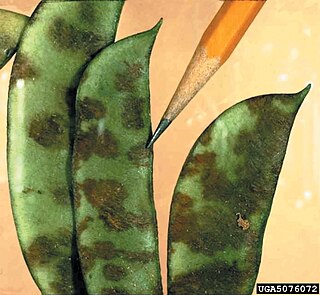
Colletotrichum lindemuthianum is a fungus which causes anthracnose, or black spot disease, of the common bean plant. It is considered a hemibiotrophic pathogen because it spends part of its infection cycle as a biotroph, living off of the host but not harming it, and the other part as a necrotroph, killing and obtaining nutrients from the host tissues.

<i>Entomophthora</i> Genus of fungi

Entomophthora is a fungal genus in the family Entomophthoraceae. Species in this genus are parasitic on flies and other two-winged insects. The genus was circumscribed by German physician Johann Baptist Georg Wolfgang Fresenius (1808–1866) in 1856.

<i>Entomophthora muscae</i> Type of pathogenic fungus

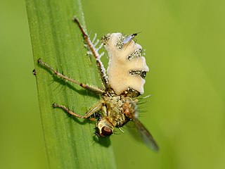
Entomophthora muscae is a species of pathogenic fungus in the order Entomophthorales which causes a fatal disease in flies. It can cause epizootic outbreaks of disease in houseflies and has been investigated as a potential biological control agent.

<i>Purpureocillium lilacinum</i> Species of fungus

Purpureocillium lilacinum is a species of filamentous fungus in the family Ophiocordycipitaceae. It has been isolated from a wide range of habitats, including cultivated and uncultivated soils, forests, grassland, deserts, estuarine sediments and sewage sludge, and insects. It has also been found in nematode eggs, and occasionally from females of root-knot and cyst nematodes. In addition, it has frequently been detected in the rhizosphere of many crops. The species can grow at a wide range of temperatures – from 8 to 38 °C for a few isolates, with optimal growth in the range 26 to 30 °C. It also has a wide pH tolerance and can grow on a variety of substrates. P. lilacinum has shown promising results for use as a biocontrol agent to control the growth of destructive root-knot nematodes.

<i>Lichtheimia corymbifera</i> Species of fungus

Lichtheimia corymbifera is a thermophilic fungus in the phylum Zygomycota. It normally lives as a saprotrophic mold, but can also be an opportunistic pathogen known to cause pulmonary, CNS, rhinocerebral, or cutaneous infections in animals and humans with impaired immunity.

<i>Actinoplanes italicus</i> Species of bacterium

Actinoplanes italicus is distinguished by the cherry-red color of its vegetative mycelium, and by the production of soluble pigments. It is also known to produce sporangia when cultured on starch or skim milk agar. Very few strains have been found and cultured, thus A. italicus is relatively uncharacterized.

<i>Rhizopus oryzae</i> Species of fungus

Rhizopus oryzae is a filamentous heterothallic microfungus that occurs as a saprotroph in soil, dung, and rotting vegetation. This species is very similar to Rhizopus stolonifer, but it can be distinguished by its smaller sporangia and air-dispersed sporangiospores. It differs from R. oligosporus and R. microsporus by its larger columellae and sporangiospores. The many strains of R. oryzae produce a wide range of enzymes such as carbohydrate digesting enzymes and polymers along with a number of organic acids, ethanol and esters giving it useful properties within the food industries, bio-diesel production, and pharmaceutical industries. It is also an opportunistic pathogen of humans causing mucormycosis.

<i>Rhizopus stolonifer</i> Species of fungus

Rhizopus stolonifer is commonly known as black bread mold. It is a member of Zygomycota and considered the most important species in the genus Rhizopus. It is one of the most common fungi in the world and has a global distribution although it is most commonly found in tropical and subtropical regions. It is a common agent of decomposition of stored foods. Like other members of the genus Rhizopus, R. stolonifer grows rapidly, mostly in indoor environments.

<i>Mucor circinelloides</i> Species of fungus

Mucor circinelloides is a dimorphic fungus belonging to the Order Mucorales. It has a worldwide distribution, found mostly in soil, dung and root vegetables. This species is described as not known to be able to produce mycotoxins, however it has been frequently reported to infect animals such as cattle and swine, as well as fowl, platypus and occasionally humans. Ketoacidotic patients are particularly at risk for infection by M. circinelloides.

<i>Alternaria brassicicola</i> Species of fungus

Alternaria brassicicola is a fungal necrotrophic plant pathogen that causes black spot disease on a wide range of hosts, particularly in the genus of Brassica, including a number of economically important crops such as cabbage, Chinese cabbage, cauliflower, oilseeds, broccoli and canola. Although mainly known as a significant plant pathogen, it also contributes to various respiratory allergic conditions such as asthma and rhinoconjunctivitis. Despite the presence of mating genes, no sexual reproductive stage has been reported for this fungus. In terms of geography, it is most likely to be found in tropical and sub-tropical regions, but also in places with high rain and humidity such as Poland. It has also been found in Taiwan and Israel. Its main mode of propagation is vegetative. The resulting conidia reside in the soil, air and water. These spores are extremely resilient and can overwinter on crop debris and overwintering herbaceous plants.

<i>Mortierella polycephala</i> Species of fungus

Mortierella polycephala is a saprotrophic fungus with a wide geographical distribution occurring in many different habitats from soil and plants to salt marshes and slate slopes. It is the type species of the genus Mortierella, and was first described in 1863 by Henri Coemans. A characteristic feature of the fungus is the presence of stylospores, which are aerial, spiny resting spores (chlamydospores).

References

  1. 1 2 3 4 5 Deepa, John; Aparna, Irodi; Joy Sarojini, Michael (2016). "Concurrent Infections of Conidiobolus Coronatus with Disseminated Tuberculosis Presenting as Bilateral Orbital Cellulitis". Journal of Clinical and Diagnostic Research. 10 (4): ND01-2. doi:10.7860/JCDR/2016/16790.7535. PMC   4866150 . PMID   27190852.
  2. 1 2 3 Emmons, Chester W.; Bridges, Charles H. (1961). "Entomophthora coronata, the Etiologic Agent of a Phycomycosis of Horses". Mycologia. 53 (3): 307–312. doi:10.2307/3756277. JSTOR   3756277.
  3. 1 2 3 4 5 6 7 8 9 10 11 12 13 14 15 16 17 18 19 20 21 22 23 Page, Robert M.; Humber, Richard A. (1973). "Phototropism in Conidiobolus coronatus". Mycologia. 65 (2): 335–354. doi:10.2307/3758106. JSTOR   3758106. PMID   4712316.
  4. 1 2 3 4 5 6 Subramanian, C; Sobel, JD (May 2011). "A case of Conidiobolus coronatus in the vagina". Medical Mycology. 49 (4): 427–9. doi: 10.3109/13693786.2010.537700 . PMID   21108542.
  5. 1 2 3 4 5 McGinnis, Michael R. (1980). Laboratory handbook of medical mycology. New York: Academic Press. ISBN   0124828507.
  6. 1 2 3 4 5 6 7 8 Rippon, John Willard (1988). Medical mycology: the pathogenic fungi and the pathogenic actinomycetes (3rd ed.). Philadelphia, PA: Saunders. ISBN   0721624448.
  7. 1 2 3 4 5 6 7 8 9 10 11 12 13 14 15 16 17 Anderson, K.H. Domsch, W. Gams, Traute-Heidi (1981). Compendium of soil fungi . London: Academic Press. ISBN   0122204018.{{cite book}}: CS1 maint: multiple names: authors list (link)
  8. 1 2 3 4 5 6 7 8 9 10 11 12 13 Kwon-Chung, K. June; Bennett, Joan E. (1992). Medical mycology. Philadelphia: Lea & Febiger. ISBN   0812114639.
  9. 1 2 3 4 5 6 Dolin, edited by Gerald L. Mandell, John E. Bennett, Raphael (2000). Mandell, Douglas, and Bennett's principles and practice of infectious diseases (5th ed.). Philadelphia: Churchill Livingstone. ISBN   044307593X.{{cite book}}: |first1= has generic name (help)CS1 maint: multiple names: authors list (link)
  10. 1 2 3 4 5 6 Reiss, Errol; Shadomy, H. Jean; Lyon, G. Marshall (2011). Fundamentals of Medical Mycology. Oxford: Wiley-Blackwell. ISBN   978-0-470-17791-4.
  11. 1 2 3 Shankar, S; Laxman, RS (Nov 2011). "Immobilization of Conidiobolus coronatus Alkaline Protease on Waste Fungal Biomass". Environmental Engineering and Management Journal. 10 (11): 1727–1732. doi:10.30638/eemj.2011.235.
  12. "Conidiobolus coronatus NRRL 28638 (ID 6815) - Genome - NCBI". www.ncbi.nlm.nih.gov.
  13. 1 2 3 Procop, Gary W.; Pritt, Bobby S. (2014). Pathology of infectious diseases. Philadelphia, PA: Elsevier Saunders. ISBN   9781437707625.
  14. Dhurat, R; Kothavade, RJ; Kumar, A (2018). "A first-line antiretroviral therapy-resistant HIV patient with rhinoentomophthoromycosis". Indian J Med Microbiol. 36 (1): 136–139. doi: 10.4103/ijmm.IJMM_16_330 . PMID   29735845.